组成图示
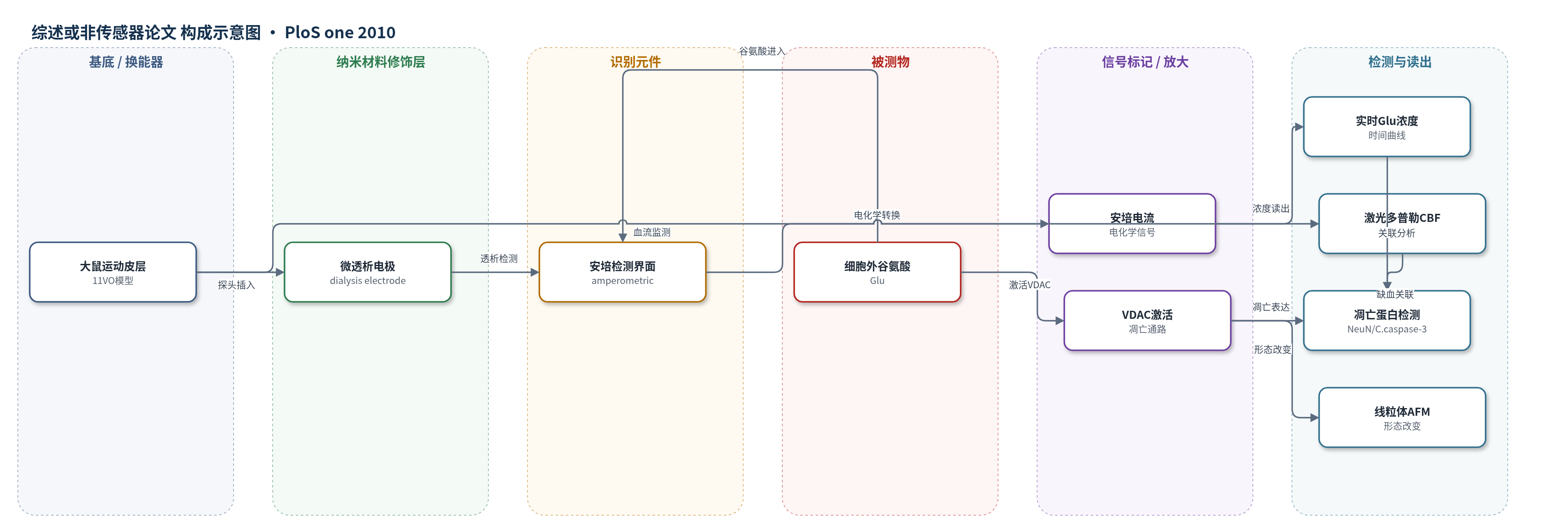
点击图片查看大图 · 依据论文自动绘制
传感器类型
综述或非传感器论文
检测对象
谷氨酸(glutamate, Glu)、脑血流(cerebral blood flow, CBF);样品基质:大鼠运动皮层细胞外液(微透析液)及在体脑组织
检测原理
在 11 血管闭塞诱导脑缺血后,神经元细胞外谷氨酸释放增加。谷氨酸经植入运动皮层的微透析电极进入透析液,由安培微透析生物传感器进行电化学检测。检测界面将谷氨酸浓度变化转换为电流信号,电流大小随谷氨酸浓度升高而增大,从而实时反映细胞外谷氨酸水平。原文未明确电极修饰酶或纳米材料,但安培法通常依赖谷氨酸或其氧化产物的电化学氧化/还原。缺血导致谷氨酸持续升高,再灌注后仍出现峰值;随后高谷氨酸与 VDAC 激活、促凋亡蛋白表达及线粒体形态改变相关联。
检测灵敏度
—
效应效果
该研究未评价传感器选择性、抗干扰、稳定性、RSD、回收率或与其他传感方法对比。其实际表现主要体现在 11VO 模型可重复性:闭塞后 %CBF 降至 12.7±2.1%,缺血平台时间 14.6±3.5 s;再灌注最大 %CBF 为 272.6±43.9%,达峰时间 691.1±246.8 s。谷氨酸释放延迟 114.9±20.2 s,缺血期最大 139.1±18.8 mM,再灌注期最大 140.7±16.7 mM。72 h 后缺血组海马 CA1 可见细胞比例 20.5±4.8%,NeuN 阳性细胞为正常组 36.0±4.2%,VDAC、cleaved caspase-3、Bax、p53 表达升高,AFM 显示线粒体表面粗糙、外膜塌陷、内膜分隔。作者主张谷氨酸释放促进 VDAC 介导凋亡。
传感器的构成
- 微透析电极(dialysis electrode):插入大鼠运动皮层,采集细胞外液并允许谷氨酸进入透析液
- 安培微透析生物传感器(amperometric microdialysis biosensor):对透析液中谷氨酸进行电化学检测
- 激光多普勒血流仪(laser-Doppler flowmetry):同步监测脑血流(CBF)变化
- 未明确识别元件:原文未报告抗体、酶、DNA探针或MIP等具体识别材料
- 未明确信号标记物:原文未报告标记物、封闭剂或电子供体
中文摘要
电压依赖性阴离子通道(VDAC)是线粒体介导凋亡的主要蛋白,其调节可能由细胞外谷氨酸过度释放诱导。本研究考察大鼠十一血管闭塞模型中谷氨酸释放对 VDAC 介导凋亡的作用。雄性 Sprague-Dawley 大鼠(250–350 g)用于 11 血管闭塞缺血模型,诱导 10 min 短暂闭塞。缺血及初始再灌注期间,使用安培微透析生物传感器实时监测细胞外谷氨酸浓度,并用激光多普勒血流仪监测脑血流(CBF)。为确认神经元凋亡,缺血 72 h 后取脑检测神经元特异性核蛋白及促凋亡蛋白(cleaved caspase-3、VDAC、p53 和 BAX),并用原子力显微镜测量线粒体形态变化。结果发现,闭塞后 CBF 百分比下降,谷氨酸释放增加,并在缺血期持续升高;缺血组谷氨酸释放水平显著更高。缺血组谷氨酸水平升高导致海马 VDAC 和促凋亡蛋白激活,并伴线粒体形态改变。研究提示,谷氨酸释放增加可促进 11 血管闭塞缺血模型中 VDAC 介导的凋亡。
英文摘要
Voltage-dependent anion channel (VDAC) is the main protein in mitochondria-mediated apoptosis, and the modulation of VDAC may be induced by the excessive release of extracellular glutamate. This study examined the role of glutamate release on VDAC-mediated apoptosis in an eleven vessel occlusion model in rats. Male Sprague-Dawley rats (250-350 g) were used for the 11 vessel occlusion ischemic model, which were induced for a 10-min transient occlusion. During the ischemic and initial reperfusion episode, the real-time monitoring of the extracellular glutamate concentration was measured using an amperometric microdialysis biosensor and the cerebral blood flow (CBF) was monitored by laser-Doppler flowmetry. To confirm neuronal apoptosis, the brains were removed 72 h after ischemia to detect the neuron-specific nuclear protein and pro-apoptotic proteins (cleaved caspase-3, VDAC, p53 and BAX). The changes in the mitochondrial morphology were measured by atomic force microscopy. A decrease in the % of CBF was observed, and an increase in glutamate release was detected after the onset of ischemia, which continued to increase during the ischemic period. A significantly higher level of glutamate release was observed in the ischemia group. The increased glutamate levels in the ischemia group resulted in the activation of VDAC and pro-apoptotic proteins in the hippocampus with morphological alterations to the mitochondria. This study suggests that an increase in glutamate release promotes VDAC-mediated apoptosis in an 11 vessel occlusion ischemic model.