组成图示
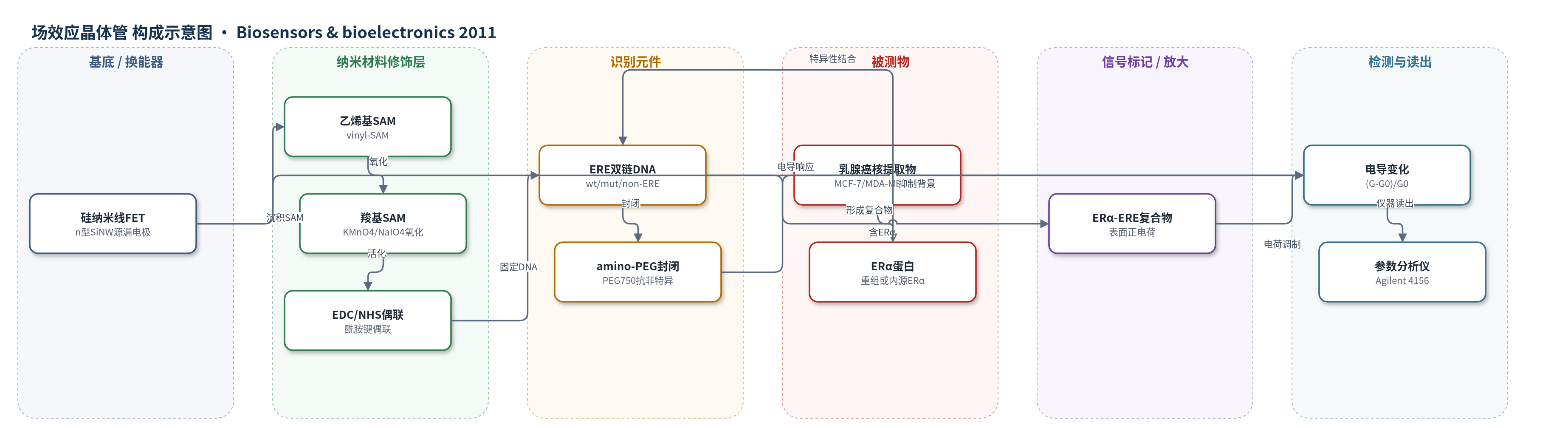
点击图片查看大图 · 依据论文自动绘制
传感器类型
场效应晶体管(FET)生物传感器
检测对象
雌激素受体α(estrogen receptor alpha, ERα)与雌激素反应元件(estrogen response element, ERE)的蛋白–DNA相互作用;样品基质:重组ERα HEPES缓冲液、乳腺癌细胞核提取物(MCF-7、MDA-MB-231)。
检测原理
该传感器以n型SiNW FET为换能器。乙烯基SAM经氧化形成羧基,通过EDC/NHS与5′-胺修饰的ERE双链DNA共价固定,amino-PEG封闭非特异位点。检测缓冲液pH 7.4低于ERα等电点(约8.3),ERα带净正电荷。当ERα与表面固定ERE特异性结合后,正电荷靠近SiNW表面,引起n型SiNW沟道电荷积累,源漏电导增加;电导变化按(G-G0)/G0读出。ERα浓度越高,结合量越多,电导增幅越大。该过程无标记、无酶放大,依靠纳米线小截面积增强表面电荷对沟道电导的调制。
检测灵敏度
LOD: 10 fM
效应效果
传感器对野生型ERE功能化表面结合ERα产生约33%电导增加,突变型约8.4%,乱序型几乎无响应,显示高序列特异性。AFM负对照中PEG封闭表面未见ERα吸附。LOD为10 fM,比SPR方法低约3个数量级。在乳腺癌细胞核提取物中,MCF-7(ER+)产生约23.4%电导变化,siRNA敲低ERα的MCF-7约5.6%,MDA-MB-231(ER-)约4.3%,表明可在复杂样品中直接检测ERα–DNA相互作用。论文未报告稳定性、重现性RSD和加标回收率。作者认为该无标记SiNW传感器可用于研究ER介导的乳腺癌基因表达。
传感器的构成
- 基底/换能器:n型硅纳米线(SiNW)场效应晶体管(FET),含源漏电极,作为电导换能器
- SAM修饰层:乙烯基终止十四烷基三氯硅烷(tetradecyltrichlorosilane)自组装单分子层(SAM),形成有序界面并钝化硅氧化层
- 氧化功能化层:KMnO4/NaIO4/K2CO3将乙烯基氧化为羧基(COOH),提供共价偶联位点
- 偶联层:EDC/NHS活化羧基,与胺修饰DNA形成酰胺键
- 识别元件:5′-胺修饰的雌激素反应元件双链DNA(ERE, dsDNA),包括wt-ERE、mut-ERE和non-ERE,特异性识别ERα
- 封闭层:氨基聚乙二醇(amino-PEG, PEG750)封闭非特异吸附
- 读出电极/仪器:源漏电极与Agilent 4156参数分析仪,测量SiNW电导变化
中文摘要
雌激素受体(ER)存在大量不同序列模式的结合位点,需要高灵敏方法区分ER–DNA结合亲和力的细微差异。本文报道了一种自组装单分子层(SAM)辅助的硅纳米线(SiNW)生物传感器,用于特异、高灵敏地检测蛋白–DNA相互作用,尤其适用于乳腺癌细胞核提取物。研究以雌激素反应元件(ERE,双链DNA)与雌激素受体α(ERα)结合为模型。SiNW表面先涂覆乙烯基终止SAM,再经氧化将末端转变为羧基,随后将胺修饰DNA固定于SiNW表面,并用该功能化传感器研究蛋白–DNA结合。X射线光电子能谱(XPS)和原子力显微镜(AFM)分别用于表征裸硅表面SAM与DNA的逐步功能化,以及可视化SiNW表面的蛋白–DNA结合。结果表明,ERα对功能化有野生型、突变型和乱序ERE的SiNW生物传感器具有高度序列特异性;该特异性DNA功能化SiNW传感器可检测低至10 fM的ERα。更重要的是,该传感器能在乳腺癌细胞核提取物中检测ERα–DNA相互作用。SAM辅助SiNW生物传感器作为无标记、高灵敏工具,在研究蛋白–DNA相互作用方面具有应用潜力。
英文摘要
The large number of estrogen receptor (ER) binding sites of various sequence patterns requires a sensitive detection to differentiate between subtle differences in ER-DNA binding affinities. A self-assembled monolayer (SAM)-assisted silicon nanowire (SiNW) biosensor for specific and highly sensitive detection of protein-DNA interactions, remarkably in nuclear extracts prepared from breast cancer cells, is presented. As a typical model, estrogen receptor element (ERE, dsDNA) and estrogen receptor alpha (ERα, protein) binding was adopted in the work. The SiNW surface was coated with a vinyl-terminated SAM, and the termination of the surface was changed to carboxylic acid via oxidation. DNA modified with amine group was subsequently immobilized on the SiNW surface. Protein-DNA binding was finally investigated by the functionalized SiNW biosensor. X-ray photoelectron spectroscopy (XPS) and atomic force microscopy (AFM) were employed to characterize the stepwise functionalization of the SAM and DNA on bare silicon surface, and to visualize protein-DNA binding on the SiNW surface, respectively. We observed that ERα had high sequence specificity to the SiNW biosensor which was functionalized with three different EREs including wild-type, mutant and scrambled DNA sequences. We also demonstrate that the specific DNA-functionalized SiNW biosensor was capable of detecting ERα as low as 10 fM. Impressively, the developed SiNW biosensor was able to detect ERα-DNA interactions in nuclear extracts from breast cancer cells. The SAM-assisted SiNW biosensor, as a label-free and highly sensitive tool, shows a potential in studying protein-DNA interactions.