组成图示
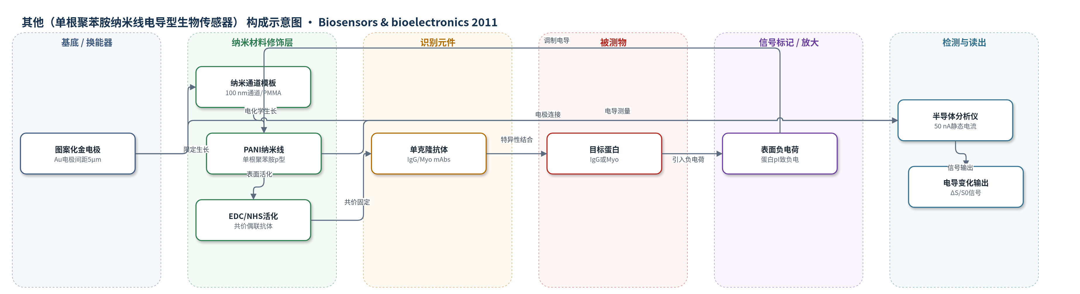
点击图片查看大图 · 依据论文自动绘制
传感器类型
其他(单根聚苯胺纳米线电导型生物传感器)
检测对象
免疫球蛋白G(immunoglobulin G, IgG)、肌红蛋白(myoglobin, Myo);样品基质:PBS缓冲液(pH 7.4)蛋白溶液,文中提及人血清应用背景
检测原理
单根PANI纳米线为p型导电聚合物,两端连接图案化Au电极。EDC/NHS将IgG或Myo单克隆抗体共价固定于纳米线表面,BSA封闭非特异性位点。目标蛋白与抗体特异性结合后,由于IgG等电点pI为7.2、Myo为6.85,在PBS pH 7.4中带弱负电荷,使纳米线表面负电荷增加。对p型PANI而言,负表面电荷引起空穴积累,电导增大。仪器施加50 nA静态电流,测量电导变化ΔS/S0。目标蛋白浓度越高,结合量越多,表面负电荷越多,电导变化越大,形成阶梯式响应。该机制为无标记电导型检测,不依赖酶催化或核酸放大,主要依靠单根纳米线高比表面积和单纳西门子级测量分辨率。
检测灵敏度
LOD: 3 ng/mL (IgG);LOD: 1.4 ng/mL (Myo);检测范围: 3 ng/mL–3 μg/mL (IgG);检测范围: 1.4 ng/mL–2.5 μg/mL (Myo)
效应效果
传感器对非特异性蛋白BSA无响应,选择性良好;非功能化纳米线对PBS、BSA(10 μg/mL)和IgG均无响应。IgG检测中,50 μg/mL抗体在10 ng/mL约产生40%电导变化,300 ng/mL–2 μg/mL饱和约50%;100 μg/mL抗体在3 ng/mL–3 μg/mL范围响应良好,3 μg/mL时电导变化可达300%;200 μg/mL灵敏度较低。Myo检测中,100 μg/mL抗体在1.4 ng/mL–2.5 μg/mL范围灵敏度和线性优于200 μg/mL。响应时间数秒,LOD为1.4 ng/mL,低于人血清正常Myo范围6–85 ng/mL,作者认为适用于心脏标志物及其他蛋白检测。
传感器的构成
- 基底/换能器电极:图案化金(Au)电极,间距5 μm,提供电学接触并测量单根纳米线电导。
- 纳米通道模板:100 nm宽纳米通道及PMMA光刻胶层,限定单根PANI纳米线原位生长,生长后去除PMMA。
- 导电聚合物纳米线:单根聚苯胺(PANI)纳米线,宽约105.4 nm,p型导电通道,作为电导换能元件。
- 交联活化层:EDC/NHS(0.2 M/0.2 M),活化表面并促进单克隆抗体与PANI共价结合。
- 识别元件:IgG或Myo单克隆抗体(mAbs,优化浓度100 μg/mL),特异性捕获目标蛋白。
- 封闭层:牛血清白蛋白(BSA,2 mg/mL),封闭非特异性蛋白结合位点。
- 信号标记物:无标记(label-free),目标蛋白负电荷改变纳米线表面电荷;Texas Red仅用于功能化验证。
中文摘要
建立了一种基于单根聚苯胺(PANI)纳米线的生物传感器,用于检测免疫球蛋白G(IgG)和肌红蛋白(Myo,心脏生物标志物之一)。单根PANI纳米线通过电化学生长法制备,在成对图案化电极之间形成单根纳米线。采用1-乙基-3-(3-二甲基氨基丙基)碳二亚胺(EDC)和N-羟基琥珀酰亚胺(NHS)辅助的表面固定法,将IgG或Myo单克隆抗体(mAbs)功能化到单根PANI纳米线上,并通过拉曼光谱和荧光显微镜验证。通过测量功能化单根PANI纳米线因mAbs捕获目标蛋白而发生的电导变化,检测IgG和Myo。IgG和Myo的检出限分别为3 ng/mL和1.4 ng/mL。当单根纳米线暴露于非特异性蛋白时未观察到响应,表明对预期目标具有优异特异性。结合快速响应时间(数秒)、高灵敏度和良好特异性,该单根PANI纳米线生物传感器在心脏标志物及其他蛋白检测中具有重要应用前景。
英文摘要
A single polyaniline (PANI) nanowire-based biosensor was established to detect immunoglobulin G (IgG) and myoglobin (Myo), which is one of the cardiac biomarkers. The single PANI nanowires were fabricated via an electrochemical growth method, in which single nanowires were formed between a pair of patterned electrodes. The single PANI nanowires were functionalized with monoclonal antibodies (mAbs) of IgG or Myo via a surface immobilization method, using 1-ethyl-3-(3-dimethyaminopropyl) carbodiimide (EDC), and N-hydroxysuccinimde (NHS). The functionalization was then verified by Raman spectroscopy and fluorescence microscopy. The target proteins of IgG and Myo were detected by measuring the conductance change of functionalized single PANI nanowires owing to the capturing of target proteins by mAbs. The detection limit was found to be 3 ng/mL for IgG and 1.4 ng/mL for Myo. No response was observed when single nanowires were exposed to a non-specific protein, demonstrating excellent specificity to expected target detection. Together with the fast response time (a few seconds), high sensitivity, and good specificity, this single PANI nanowire-based biosensor shows great promise in the detection of cardiac markers and other proteins.