组成图示
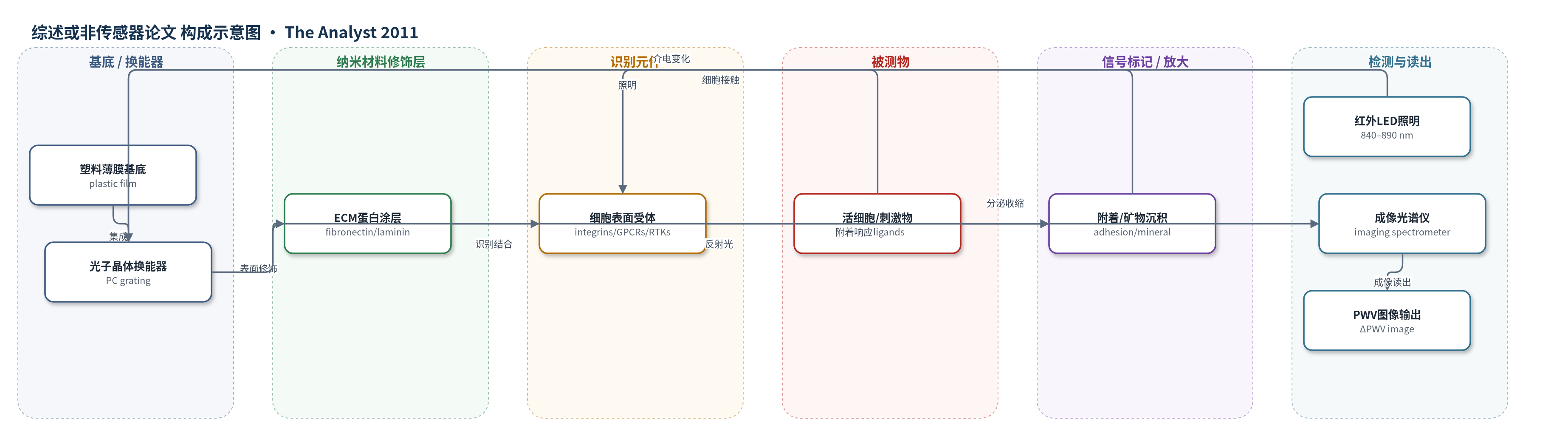
点击图片查看大图 · 依据论文自动绘制
传感器类型
综述或非传感器论文
检测对象
活细胞(cells,如HEK293、CHO、HeLa、A431、MSC、心肌细胞、肝细胞)及其对配体/化合物(agonists/antagonists/toxins/chemokines)的响应;样品基质:细胞培养液/微孔板培养体系。
检测原理
光子晶体(PC)由周期性介电结构构成,在特定波长产生共振反射。当细胞或生物分子吸附于PC表面时,表面附近介电常数改变,使反射峰波长(PWV)发生位移;细胞数量、黏附强度、形态收缩、受体激活或矿物沉积越多,PWV位移越大。检测时从微孔板下方用红外LED照明,反射光由光谱仪或成像光谱仪采集,软件拟合得到PWV或逐像素PWV图像。通过前后图像相减和负对照扣除缓冲液折射率影响,可实时监测细胞附着、受体激活、细胞毒性、趋化和干细胞分化。该体系无外源标记,主要依靠细胞群体响应或成骨矿物沉积累积产生信号,成像掩膜算法可提高低细胞数信噪比。
检测灵敏度
未报告LOD、线性范围、灵敏度斜率或R^2。
效应效果
该综述强调PC光学生物传感器为无标记、可重复、可长期监测的细胞检测平台。传感器集成于96/384/1536孔微孔板,Reader采用8个检测头,单点直径约0.6 mm,8探针采集约20 ms,96孔约5 s;Scanner成像分辨率可达3.75×3.75 μm²,并用掩膜算法将384孔低细胞数检测降至约250 cells/well,某些实验低于100 cells/well。GPCR、RTK和TRPV1激活产生不同动力学PWV,capsaicin可诱导约1000 pm负移,TRPV1拮抗剂IC50与正交测量呈1:1线性关系。细胞毒性按机制呈现不同profile;心肌细胞搏动可测频率与振幅;MSC成骨分化PWV位移大于30 nm,比Alizarin Red染色早3–4天。作者认为其适用于药物高通量筛选。
传感器的构成
- 基底/换能器:塑料薄膜(plastic film)上复制成型的光子晶体(PC)周期介电结构,作为光学换能器,反射峰波长(PWV)随表面介电变化。
- 微孔板载体:标准96/384/1536孔塑料微孔板(microplate),PC集成于孔底,兼容细胞培养与液体处理。
- 表面功能化层:细胞外基质(ECM)蛋白如纤连蛋白(fibronectin)、层粘连蛋白(laminin)、胶原(collagen)、多聚赖氨酸(poly-D-lysine),促进细胞附着。
- 识别元件:细胞表面受体/整合素(integrins,如α4β1/VLA-4)或表面捕获的细胞黏附分子(CAM,如VCAM);也可用抗体/肽(antibodies/peptides)选择性捕获细胞。
- 信号标记物:无外源标记(label-free),活细胞附着、收缩或矿物沉积改变局部介电,产生PWV位移。
- 读出层:红外LED(840–890 nm)从板下照明,反射光由光谱仪/成像光谱仪(spectrometer)采集,得到PWV或PWV图像。
中文摘要
本文综述了光子晶体(PC)光学生物传感器在多种无标记细胞检测中的应用。PC由周期性介电结构组成,可反射特定波长;当细胞或生物分子吸附于传感器表面时,局部介电常数改变,使反射峰波长(PWV)发生位移,从而无需染料即可定量监测细胞附着、增殖、凋亡、细胞毒性、趋化、离子通道激活及膜蛋白激活等过程。与显微镜相比,该传感器可监测整合素—表面相互作用的动态变化,且不受光漂白影响。文中介绍了集成于96、384或1536孔微孔板的PC传感器,以及两种读出仪器:BIND Reader用于群体细胞附着测量,BIND Scanner用于单细胞成像,空间分辨率可达3.75×3.75 μm²。作者还讨论了GPCR、受体酪氨酸激酶、离子通道、细胞黏附、细胞毒性、低细胞数检测、异质细胞群、干细胞分化和趋化等应用,强调其在药物高通量筛选和生物相关细胞检测中的价值。
英文摘要
Biosensor technologies that have been primarily used in the past for characterizing biomolecular interactions are now being used to develop new approaches for performing cell-based assays. Biosensors monitor cell attachment to a transducer surface, and thus provide information that is fundamentally different from that provided by microscopy, as the sensor is capable of monitoring temporal evolution of integrin-surface interactions that are difficult to measure by other means. Label-free biosensor technologies are especially advantageous for monitoring the behavior of cells because they do not require stains that typically result in cell death, and are not subject to effects such as photobleaching. As a result, cells can be quantitatively monitored in their culture environment over an extended period of time while processes such as proliferation, apoptosis, cytotoxicity, chemotaxis, ion channel activation, and membrane-bound protein activation are modulated by the introduction of a variety of chemical or biological stimuli. This review describes the application of photonic crystal optical biosensor microplates to a variety of cell-based assays. Detection instruments for photonic crystals measure the aggregate behavior of large cell populations, or, using recently developed biosensor imaging detection, independent monitoring of individual cells. These technological developments offer the ability to perform assays with a limited number of available cells for applications such as high throughput screening with primary cells or stem cells.