组成图示
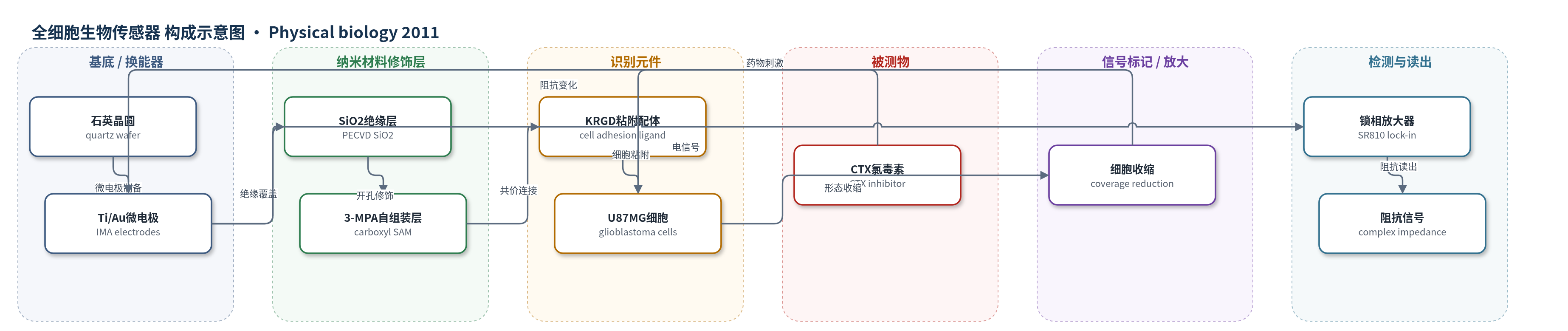
点击图片查看大图 · 依据论文自动绘制
传感器类型
全细胞生物传感器
检测对象
氯毒素(chlorotoxin, CTX);样品基质:O-MEM 培养基中贴壁 U87MG 人胶质母细胞瘤细胞
检测原理
本传感器以贴附在微电极上的活细胞作为识别元件。CTX 进入细胞并调节氯离子通道介导的体积调控,使原本铺展的 U87MG 细胞收缩、变圆,细胞-电极覆盖面积 Ac 减小,细胞-电极缝隙暴露。交流电流主要沿细胞旁路(paracellular)通过电解质,覆盖面积变化改变电极-电解质界面阻抗 Ze,free 和密封电阻 Rseal,使整体复阻抗 Z 的实部下降。锁相放大器在 2 kHz 施加 10 mV 正弦波,测量电压幅值和相位并计算复阻抗。CTX 浓度越高,细胞收缩越明显,实部阻抗下降越大;500 nM 基本无变化,5 和 10 μM 约下降 50%。单细胞电极减少旁路电流和细胞-电极间距,提高信噪比。
检测灵敏度
原文未报告 LOD、线性范围、灵敏度斜率或相关系数。
效应效果
系统通过单细胞电极显著提高细胞信号:单细胞电极最大细胞-基线比 CBR 为 1.59,多细胞电极仅 0.62;2 kHz 下 CBR 较 500 Hz 提高 9 倍、较 20 kHz 提高 3 倍。100 nA 交流刺激 24 h 后细胞存活率为 100%,100 μA 仅 3.8%,说明低电流可保持细胞活性。5 μM CTX 处理 16 h,8 h 后四个单细胞实部阻抗均下降 50% 以上,而多细胞电极几乎无变化,无 CTX 对照稳定。500 nM CTX 基本无响应,1 μM 仅部分细胞下降,5 和 10 μM 响应相近,提示 5 μM 为有效剂量。作者认为该系统实时、无标记、高通量且可寻址,可用于药物筛选、个性化医疗和环境毒素检测。
传感器的构成
- 基底/换能器电极:石英晶圆(quartz wafer)上 Ti/Au 微电极阵列(IMA),含 20/30 μm 单细胞检测电极、250 μm 多细胞检测电极和矩形对电极,Ti 增强粘附、Au 导电。
- 绝缘钝化层:PECVD SiO2(约 1 μm)覆盖互连并开孔暴露电极,提供电绝缘。
- 电极表面修饰层:3-巯基丙酸(3-MPA)自组装单分子层(SAM),提供羧基用于共价连接细胞粘附配体。
- 非电极区钝化层:PEG 硅烷(2-[methoxy(polyethyleneoxy)propyl] trimethoxysilane)钝化 SiO2/硅背景,抗蛋白吸附和细胞附着。
- 识别元件:KRGD 寡肽通过 EDAC/NHS 活化 3-MPA 羧基共价连接,介导 U87MG 细胞天然粘附与铺展。
- 被测细胞:U87MG 人胶质母细胞瘤细胞,在 O-MEM 培养基中贴附于电极,其形态和阻抗变化被检测。
- 读出电路:MUX/预放大 PCB、SR810 锁相放大器、LabVIEW/MATLAB,施加 10 mV 正弦波并测量电压幅值与相位以计算复阻抗。
中文摘要
本文报道了一种用于实时监测细胞对化学或生物试剂响应的芯片式细胞阻抗生物传感器。细胞阻抗变化可为生物医学和环境应用提供有用信息,而基于单细胞的分析平台有望获得更灵敏的细胞阻抗测量,尤其适用于预期阻抗变化微小的场景。作者开发了具有两类电极的芯片细胞阻抗传感器,分别承载单个细胞和细胞群,以评估其检测细胞响应的效能。人胶质母细胞瘤U87MG细胞通过配体介导的天然细胞粘附被图案化在单细胞和多细胞电极上。研究比较了这两种电极上的癌细胞对离子通道抑制剂氯毒素(CTX)的形态变化和阻抗变化,以利用单细胞系统的精细检测能力。承载单个细胞的检测电极表现出实部阻抗信号显著降低,而承载致密单层细胞的电极几乎无阻抗变化。用不同剂量CTX处理单细胞电极时,观察到剂量依赖的阻抗变化,从而可识别该治疗所需的有效剂量。研究证明,该单细胞阻抗系统可作为环境毒素检测和药物评价等生物医学应用的有用分析工具。
英文摘要
The response of cells to a chemical or biological agent in terms of their impedance changes in real-time is a useful mechanism that can be utilized for a wide variety of biomedical and environmental applications. The use of a single-cell-based analytical platform could be an effective approach to acquiring more sensitive cell impedance measurements, particularly in applications where only diminutive changes in impedance are expected. Here, we report the development of an on-chip cell impedance biosensor with two types of electrodes that host individual cells and cell populations, respectively, to study its efficacy in detecting cellular response. Human glioblastoma (U87MG) cells were patterned on single- and multi-cell electrodes through ligand-mediated natural cell adhesion. We comparatively investigated how these cancer cells on both types of electrodes respond to an ion channel inhibitor, chlorotoxin (CTX), in terms of their shape alternations and impedance changes to exploit the fine detectability of the single-cell-based system. The detecting electrodes hosting single cells exhibited a significant reduction in the real impedance signal, while electrodes hosting confluent monolayer of cells showed little to no impedance change. When single-cell electrodes were treated with CTX of different doses, a dose-dependent impedance change was observed. This enables us to identify the effective dose needed for this particular treatment. Our study demonstrated that this single-cell impedance system may potentially serve as a useful analytical tool for biomedical applications such as environmental toxin detection and drug evaluation.