组成图示
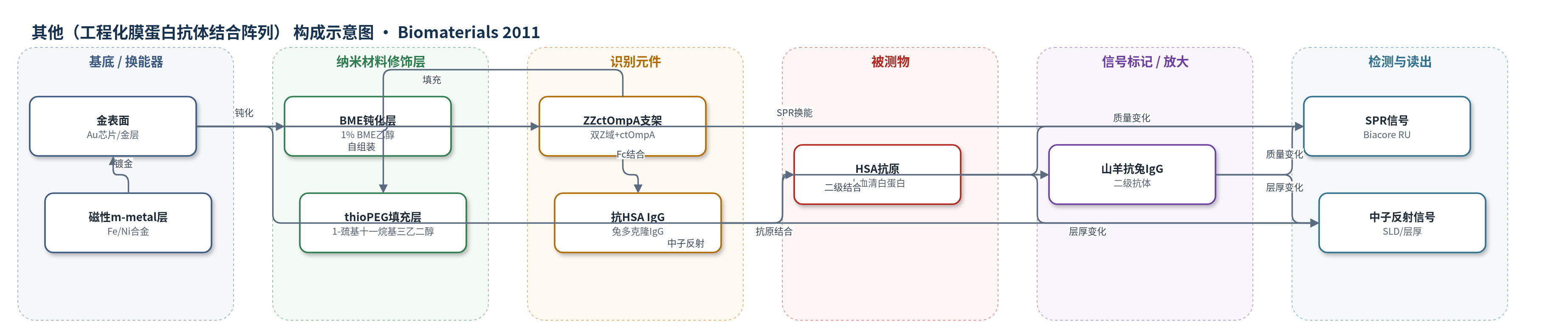
点击图片查看大图 · 依据论文自动绘制
传感器类型
其他(工程化膜蛋白抗体结合阵列)
检测对象
人血清白蛋白(HSA,human serum albumin),样品基质:PBS 缓冲液/氘代 PBS(D2O)
检测原理
ZZctOmpA 通过 ctOmpA 支架中的单个半胱氨酸与金表面形成硫醇-金键,经 thioPEG 填充后呈垂直取向;其 N 端两个 Z 域特异性结合 IgG 的 Fc 恒定区,使兔多克隆抗 HSA IgG 以直立方式固定,可变区朝向溶液。当 HSA 进入流动池时,与 IgG 可变区结合,界面质量与折射率增加,SPR 响应单位(RU)随抗原浓度升高并趋于饱和;加入山羊抗兔 IgG 后形成完整免疫层,进一步增加 SPR 信号。中子反射利用磁性 m-metal 层和 D2O/氘代 PBS 同位素对比,获得散射长度密度(SLD)剖面,定量 IgG、HSA 和二级抗体层厚,从而验证抗体直立取向。
检测灵敏度
原文未报告 LOD、线性范围、灵敏度斜率或 R^2。
效应效果
ZZctOmpA 阵列对兔多克隆抗 HSA IgG 的表观 Kd 为 0.137 nM,koff 2.74×10^-5 s^-1,半衰期超过 24 h;小鼠 IgG2a 的 Kd 为 0.778 nM,解离更快。ctOmpA 对照显示高浓度 IgG 下非特异结合约为 ZZctOmpA 的 20%,低浓度下无显著差异,thioPEG 表面无结合。SPR 组装响应 1000–1800 RU,对应 1.74–3.14×10^10 个蛋白/mm²;中子反射显示蛋白覆盖 15–20%(最高 40%),thioPEG 覆盖 63–72%。抗体层厚 126 Å,加 HSA 后 163 Å,加二级抗体后 210 Å,完整层约 223 Å,IgG 体积分数约 0.28,呈密集直立取向。作者认为该表面可为高灵敏度免疫诊断装置提供基础。
传感器的构成
- 基底/换能器电极:金表面(Au)/金层(Au),SPR 芯片与中子反射样品,提供硫醇-金键结合和光学/中子换能界面
- 磁性对比层:m-metal(Fe/Ni 合金,含 Cu/Mo),中子反射样品中用于磁性对比,增强 SLD 剖面分辨
- 钝化层:BME(β-巯基乙醇,1% 乙醇溶液),在金表面形成自组装层,减少非特异结合
- 填充/封闭层:thioPEG(1-巯基十一烷基三乙二醇,1-mercaptoundec-11-yltriethylene glycol),填充蛋白间隙,稳定取向并降低非特异吸附
- 识别支架:ZZctOmpA(双 Z 域融合循环置换 ctOmpA 支架),经半胱氨酸硫醇-金键取向固定,Z 域结合 IgG Fc 恒定区
- 识别元件:兔多克隆抗 HSA IgG(rabbit polyclonal anti-HSA IgG),Fc 结合 Z 域,可变区朝外结合抗原
- 信号标记/二级抗体:山羊抗兔 IgG(anti-rabbit IgG produced in goat),结合兔 IgG,用于完整免疫层和 SPR/中子反射信号
中文摘要
本文报道了一种膜蛋白阵列,可在结合免疫球蛋白 G(IgG)恒定区的同时,使可变区保持自由以结合抗原。该阵列支架为大肠杆菌外膜蛋白 A(OmpA)的跨膜域 tOmpA,经工程化改造后通过单个半胱氨酸残基在金表面组装成取向单层;其他蛋白结构域可融合到支架的 N 端和 C 端。本研究采用循环置换的 ctOmpA 融合两个金黄色葡萄球菌蛋白 A 的 Z 域(ZZctOmpA),构建 IgG 结合阵列。利用圆二色光谱评估工程蛋白的溶液结构,采用表面等离子共振(SPR)和中子反射测量阵列组装、抗体结合及抗原结合。与小鼠 IgG2 相比,兔多克隆 IgG 与 ZZctOmpA 结合更强,且免疫球蛋白解离足够慢,可支持对含抗原组装层的中子反射研究。结合磁性与同位素对比,建立了完整的逐层模型,结果显示约 223 Å 高的层中包含直立取向的抗体。
英文摘要
This paper describes a membrane protein array that binds immunoglobulin G at its constant regions whilst leaving the variable regions free to bind antigen. The scaffold of the array is the transmembrane domain of outer membrane protein A (tOmpA) from Escherichia coli engineered to assemble as an oriented monolayer on gold surfaces via a single cysteine residue. Other protein domains can be fused to the N and C termini of the scaffold. In this study we use circularly permuted ctOmpA fused to two Z domains of Staphylococcus aureus protein A (ZZctOmpA) to create the immunoglobulin G-binding array. The solution structure of the engineered proteins was assessed by circular dichroism spectroscopy. Assembly of the array, attachment of antibodies and antigen binding were measured using surface plasmon resonance and neutron reflection. Compared to mouse IgG2, polyclonal IgG from rabbit bound very strongly to ZZctOmpA and the dissociation of the immunoglobulin was slow enough to allow neutron reflection studies of the assembled layer with antigen. Using both magnetic and isotopic contrasts a complete layer by layer model was defined which revealed that the 223 Å high layer contains antibodies in an upright orientation.