组成图示
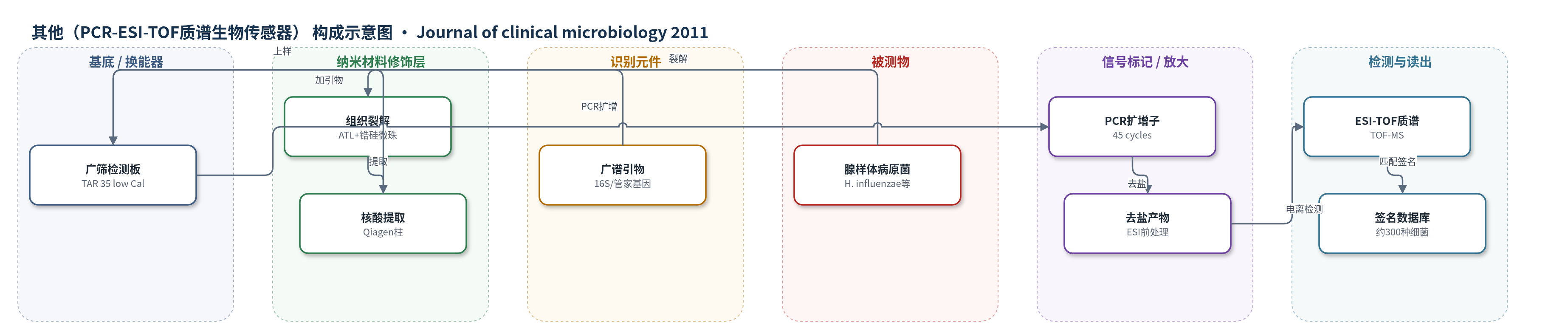
点击图片查看大图 · 依据论文自动绘制
传感器类型
其他(PCR-ESI-TOF质谱生物传感器)
检测对象
流感嗜血杆菌(Haemophilus influenzae)、肺炎链球菌(Streptococcus pneumoniae)、卡他莫拉菌(Moraxella catarrhalis)、金黄色葡萄球菌(Staphylococcus aureus)及其他上呼吸道病原菌;样品基质:儿童腺样体活检组织(adenoid biopsy tissue)
检测原理
Ibis T5000系统首先从腺样体组织中裂解并提取核酸,随后加入16种广谱细菌引物,包括16S rDNA和管家基因引物,对样本中多种保守基因进行PCR扩增。该过程不依赖预设单一病原体,而是通过扩增子质量/碱基组成形成微生物签名。PCR产物经去盐后用于电喷雾电离(ESI)飞行时间质谱(TOF-MS)分析,质谱峰及扩增子重量被用于计算碱基组成签名,并与约300种细菌数据库匹配。目标菌DNA含量越高,相应扩增子信号越强,从而反映该菌在样本中的存在与相对丰度。
检测灵敏度
原文未报告LOD、线性范围、灵敏度斜率或相关系数。
效应效果
Ibis T5000在COM组检出24种细菌,OSA组为10种,COM组多样性约为OSA组2倍。COM组流感嗜血杆菌检出率66.7%,OSA组10%,差异显著(P=0.005);肺炎链球菌两组均50%,金黄色葡萄球菌COM组8.3%、OSA组30%,无显著差异。FISH/CLSM证实病原菌聚集、被碳水化合物基质包绕并位于上皮表面或细胞内,符合生物膜标准。体外2 mg/ml阿奇霉素处理2 h后仍见活菌,提示生物膜菌耐受约100倍MIC90抗生素。PCR确认12例FISH阳性COM样本中11例含H. influenzae、11例中10例含S. pneumoniae,相关性约90%。
传感器的构成
- 样品前处理层:HBSS、ATL裂解缓冲液、0.1 mm锆硅微珠(zirconia/silica beads)与Qiagen组织研磨柱,用于破碎腺样体组织并释放核酸。
- 核酸纯化层:Qiagen mini-spin column,用于洗脱获得200 μL纯化基因组材料。
- 识别元件:16种广谱细菌引物(16S rDNA及管家基因引物组合),用于无预设靶标扩增保守基因。
- 反应/校准层:TAR 35 lower-calibrant-level(low Cal)广筛板,承载PCR反应与低校准水平检测。
- 信号标记/放大元件:PCR扩增子(amplicons),经45个循环扩增后作为质谱分析对象。
- 换能/读出层:Ibis T5000 Universal Biosensor System,通过ESI-TOF MS检测扩增子碱基组成并匹配约300种细菌数据库。
中文摘要
儿童慢性中耳炎(COM)中耳黏膜上存在病原菌生物膜,可能促进病原体持续存在并导致抗生素治疗困难。腺样体切除术对COM有效,提示腺样体可能是COM病原体的储库。本研究从35名因COM或阻塞性睡眠呼吸暂停(OSA)接受腺样体切除术的儿童中获取腺样体样本,采用一种新型无培养分子诊断方法,并结合共聚焦显微镜,原位分析腺样体中病原菌的分布与组织结构,以判断其是否符合生物膜特征。使用Ibis T5000通用生物传感器系统检测腺样体活检标本中的微生物多样性。通过16种广谱细菌引物,结果显示两组患者的腺样体均被多菌生物膜定植。COM组腺样体中流感嗜血杆菌(H. influenzae)检出率更高(P=0.005),而肺炎链球菌(S. pneumoniae)和金黄色葡萄球菌(S. aureus)在两组间无显著差异。荧光原位杂交(FISH)、凝集素结合及针对宿主上皮细胞的抗体染色显示,病原菌呈聚集状,被碳水化合物基质包绕,并定位于上皮细胞表面及细胞内,符合细菌生物膜标准。
英文摘要
Biofilms of pathogenic bacteria are present on the middle ear mucosa of children with chronic otitis media (COM) and may contribute to the persistence of pathogens and the recalcitrance of COM to antibiotic treatment. Controlled studies indicate that adenoidectomy is effective in the treatment of COM, suggesting that the adenoids may act as a reservoir for COM pathogens. To investigate the bacterial community in the adenoid, samples were obtained from 35 children undergoing adenoidectomy for chronic OM or obstructive sleep apnea. We used a novel, culture-independent molecular diagnostic methodology, followed by confocal microscopy, to investigate the in situ distribution and organization of pathogens in the adenoids to determine whether pathogenic bacteria exhibited criteria characteristic of biofilms. The Ibis T5000 Universal Biosensor System was used to interrogate the extent of the microbial diversity within adenoid biopsy specimens. Using a suite of 16 broad-range bacterial primers, we demonstrated that adenoids from both diagnostic groups were colonized with polymicrobial biofilms. Haemophilus influenzae was present in more adenoids from the COM group (P = 0.005), but there was no significant difference between the two patient groups for Streptococcus pneumoniae or Staphylococcus aureus. Fluorescence in situ hybridization, lectin binding, and the use of antibodies specific for host epithelial cells demonstrated that pathogens were aggregated, surrounded by a carbohydrate matrix, and localized on and within the epithelial cell surface, which is consistent with criteria for bacterial biofilms.