组成图示
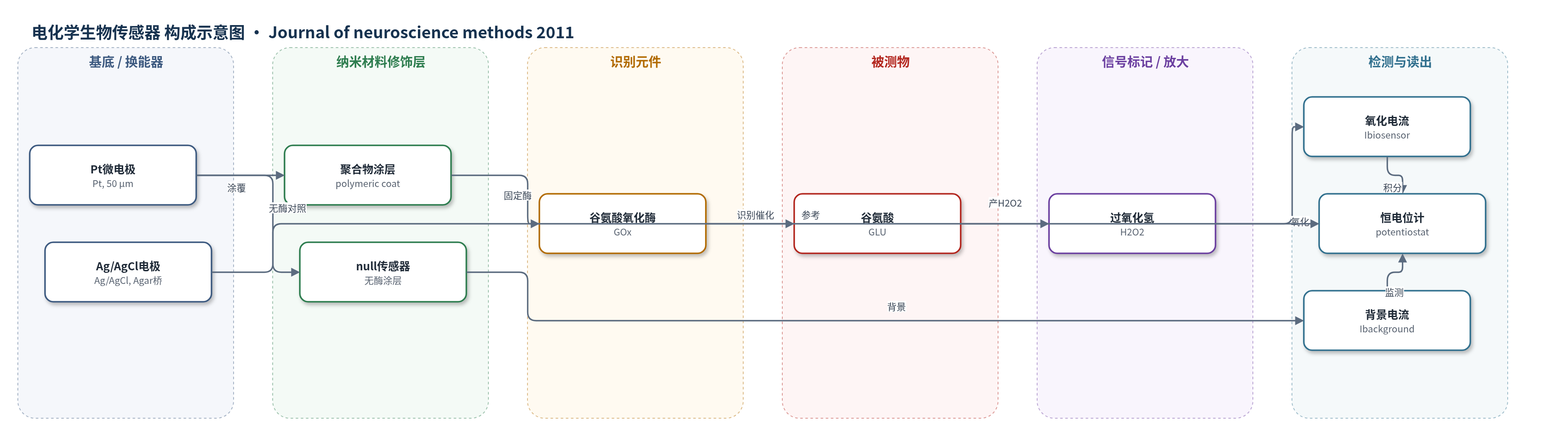
点击图片查看大图 · 依据论文自动绘制
传感器类型
电化学生物传感器
检测对象
内源性谷氨酸(glutamate, GLU);样品基质:大鼠脊髓器官型切片(organotypic slices)基础溶液
检测原理
谷氨酸氧化酶(GOx)特异性识别并催化内源性谷氨酸(GLU),生成2-oxoglutarate、NH3和H2O2。产生的H2O2扩散至Pt微电极表面,在恒电位计施加的500 mV电位下经Fenton反应被氧化,释放电子并形成氧化电流。该电流与GLU浓度呈线性关系,因此可通过校准曲线将电流换算为谷氨酸浓度。null传感器仅含聚合物涂层,用于监测非特异性电活性干扰;当背景电流稳定时,总电流扣除背景后反映GLU释放。该方法无额外化学放大,主要依赖酶催化转化与电化学氧化实现实时检测。
检测灵敏度
灵敏度: 0.5 nA/μM;线性范围: 0.1–100 μM(体外报告);校准范围: 0.5–50 μM;r = 0.9902(谷氨酸校准);r = 0.9987(H2O2校准)
效应效果
传感器对谷氨酸具有酶特异性,null传感器背景稳定(0.33±0.10、0.39±0.12、0.29±0.08 nA),未扣除。80 min内10 μM GLU响应保持起始的71.8±2.9%。0.5 mM KA使GLU峰电流1.27±0.55 nA,估算峰浓度3.10±2.05 μM,7.07±1.86 min达峰;0.1 mM KA为0.51±0.37 nA、1.66±0.37 μM。KA后坏死核约10%,NeuN神经元无显著丢失。相比微透析-HPLC,该方法可实时监测,可用于筛选靶向谷氨酸释放的神经保护药物。
传感器的构成
- 换能器电极:Pt微电极(直径50 μm、长1 mm),作为电子传导与氧化电流记录基底
- 识别/催化层:谷氨酸氧化酶(glutamate oxidase, GOx),特异性催化谷氨酸(GLU)生成2-oxoglutarate、NH3和H2O2
- 聚合物涂层:polymeric coat,用于固定酶;null传感器仅含该层作对照
- 对照传感器:null biosensor(无酶聚合物涂层),用于监测非特异性电活性干扰
- 参考电极:Ag/AgCl线,经Agar桥连接浴液,提供稳定电位参考
- 信号读出装置:多通道恒电位计(multichannel potentiostat),在500 mV下积分氧化电流
中文摘要
谷氨酸过度释放被认为是急性脊髓损伤相关兴奋毒性导致细胞损伤的主要成分。本研究利用海人酸(kainate, KA)诱导的大鼠脊髓器官型切片培养体系作为兴奋毒性体外模型,将商用谷氨酸生物传感器置于切片腹角区域,实时监测KA应用1 h后内源性谷氨酸释放的时间过程与幅度。结果显示,0.5 mM KA处理后谷氨酸释放于给药后约7 min达到峰值,随后缓慢下降,在洗脱前恢复至基线;较低浓度0.1 mM KA引起较小且发展较慢的释放。实验结束后通过计数固缩核评估细胞死亡,发现死亡细胞约占总体细胞的10%,且未见显著神经元丢失,这与既往研究提示KA诱导的神经元死亡存在延迟一致。本研究证明,谷氨酸生物传感器可用于实时监测急性脊髓损伤体外模型中内源性谷氨酸释放,为研究靶向谷氨酸释放机制的神经保护药物作用提供方法学工具。
英文摘要
Excessive release of glutamate is believed to be a major component of cell damage following excitotoxicity associated with acute spinal cord injury. Using an in vitro model of excitotoxicity evoked by kainate on rat organotypic spinal slice cultures, we investigated the timecourse and extent of endogenous glutamate release following 1h application of kainate using a commercially available biosensor placed on the ventral horn area of such slices. The release of glutamate peaked 7 min from the start of kainate (0.5 mM) application and then slowly declined to baseline prior to kainate washout. A lower concentration of kainate (0.1 mM) induced a smaller release that developed more slowly. At the end of each experiment, the number of pyknotic nuclei was counted to quantify cell death that was found to be about 10% of the total population with no significant neuronal loss. This finding accords with previous studies showing that, on the basis of neuronal counts at various times after kainate application, neuronal death was delayed. The present data demonstrate that a glutamate biosensor can be employed for real-time monitoring of endogenous glutamate release from an in vitro model of acute spinal cord injury applied to organotypic slices. This method can, therefore, be useful to study the cellular action of neuroprotective drugs targeting glutamate release mechanisms.