组成图示
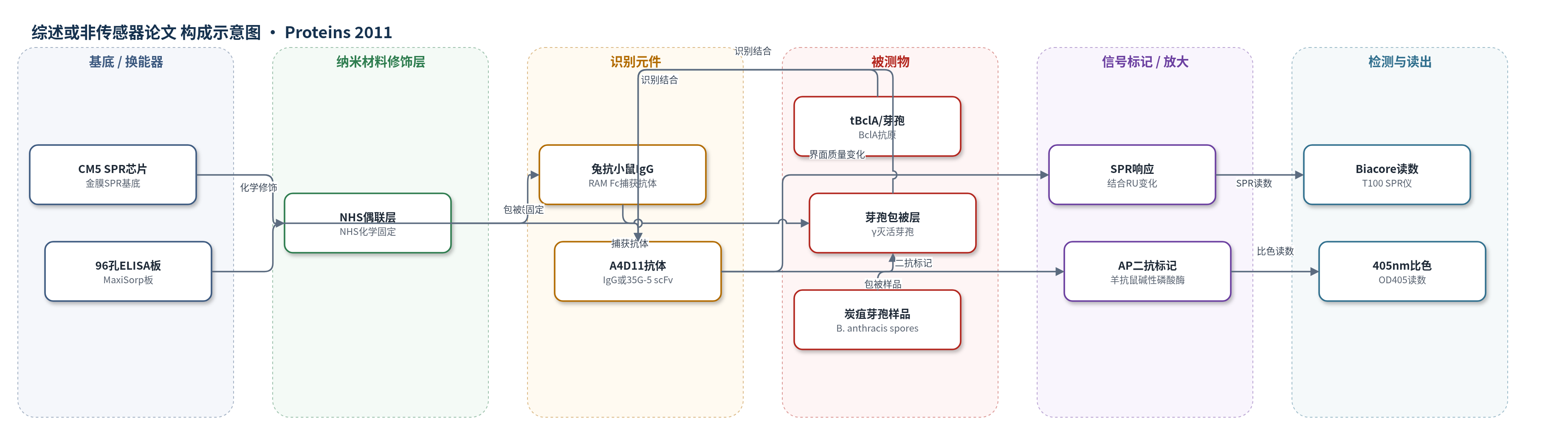
点击图片查看大图 · 依据论文自动绘制
传感器类型
综述或非传感器论文
检测对象
炭疽芽孢(Bacillus anthracis spores,γ灭活芽孢悬液)、截短BclA蛋白(tBclA,重组蛋白溶液)
检测原理
本文采用SPR和ELISA两条检测路径。SPR中,A4D11 IgG或35G-5 scFv与tBclA发生特异性结合,结合事件改变CM5金膜表面的质量/折射率,引起表面等离子共振角位移,以响应单位(RU)读出;通过不同浓度分析物的结合/解离曲线拟合1:1模型,获得ka、kd和KD,信号随结合量增加而增大。ELISA中,γ灭活芽孢包被于96孔板,A4D11或scFv识别芽孢表面BclA,羊抗鼠碱性磷酸酶二抗结合一抗并催化底物显色,405 nm吸光度与结合量正相关。未使用HCR、RCA或CRISPR等核酸放大策略,主要依赖抗体识别和酶标二抗信号放大。
检测灵敏度
—
效应效果
A4D11可识别14株γ灭活炭疽芽孢,但不能区分B. mycoides、B. thuringiensis和B. cereus;商业S26在该ELISA中特异性和反应性更高。35G-5 scFv单体/二体对芽孢敏感性低于亲代IgG,二体因亲合力优于单体,但交叉反应模式相同。SPR显示IgG KD为3.3×10^-10 M,scFv KD为2.6×10^-10 M和2.3×10^-10 M。结构表明抗体结合tBclA三聚体侧面,该表位在天然外膜中被相邻三聚体遮挡且残基保守,解释低芽孢结合与交叉反应。作者认为可经结构指导改造,用作炭疽生物传感器前端识别试剂,但现场检测仍需PCR确认。
传感器的构成
- SPR换能基底:CM5金膜SPR芯片,提供表面等离子共振检测界面
- 化学偶联层:NHS化学(EDC/NHS)用于固定捕获抗体或抗原
- 捕获/识别层:兔抗小鼠IgG(RAM Fc)固定于芯片,用于捕获A4D11 IgG;或tBclA固定于芯片作为配体
- 识别元件:A4D11单克隆抗体或35G-5 scFv,识别tBclA/芽孢BclA
- 分析物:重组tBclA蛋白溶液或γ灭活完整芽孢悬液
- 运行缓冲液:HBS-EP1(10 mM Hepes、150 mM NaCl、3 mM EDTA、0.05% P20),SPR结合环境
- ELISA固相载体:MaxiSorp 96孔板,包被γ灭活芽孢
- 封闭剂:脱脂奶,用于ELISA非特异性封闭
- 信号标记物:羊抗鼠碱性磷酸酶二抗,用于ELISA酶标显色
中文摘要
实验室或现场检测炭疽的一种方法,是利用高亲和力和高特异性结合试剂检测炭疽芽孢杆菌(Bacillus anthracis)芽孢。作者从单克隆抗体库中筛选出对炭疽芽孢外膜蛋白BclA截短形式tBclA具有高亲和力的候选抗体A4D11。利用Biacore表面等离子共振生物传感器进行标准动力学和动力学滴定分析,显示该抗体对重组tBclA的亲和力处于300 pM量级;将A4D11改造为单链可变片段(scFv)后,其亲和力未明显下降。然而,针对炭疽芽孢及近缘芽孢杆菌属物种的试验显示,该抗体对完整芽孢的结合有限,并在不同芽孢杆菌属物种间存在明显交叉反应。scFv-tBclA复合物的三维晶体结构解释了上述结果:A4D11结合tBclA三聚体的侧面,接触的是天然外膜中通常与相邻三聚体紧密堆积的抗原表面;该界面在芽孢杆菌属物种间高度保守。结果表明,针对高度保守的芽孢结构获得能区分近缘物种的高亲和力抗体较为困难,但也提示未来可通过结构指导的抗体设计改进该检测识别元件。
英文摘要
One method of laboratory- or field-based testing for anthrax is detection of Bacillus anthracis spores by high-affinity, high specificity binding reagents. From a pool of monoclonal antibodies, we selected one such candidate (A4D11) with high affinity for tBclA, a truncated version of the B. anthracis exosporium protein BclA. Kinetic analysis utilising both standard and kinetic titration on a Biacore biosensor indicated antibody affinities in the 300 pM range for recombinant tBclA, and the A4D11 antibody was also re-formatted into scFv configuration with no loss of affinity. However, assays against B. anthracis and related Bacilli species showed limited binding of intact spores as well as significant cross-reactivity between species. These results were rationalized by determination of the three-dimensional crystallographic structure of the scFv-tBclA complex. A4D11 binds the side of the tBclA trimer, contacting a face of the antigen normally packed against adjacent trimers within the exosporium structure; this inter-spore interface is highly conserved between Bacilli species. Our results indicate the difficulty of generating a high-affinity antibody to differentiate between the highly conserved spore structures of closely related species, but suggest the possibility of future structure-based antibody design for this difficult target.