组成图示
点击图片查看大图 · 依据论文自动绘制
传感器类型
化学发光生物传感器
检测对象
细胞凋亡(apoptosis)、caspase-3 激活(caspase-3 activation);样品基质:细胞条件培养基、小鼠血液、肿瘤组织/活体小鼠
检测原理
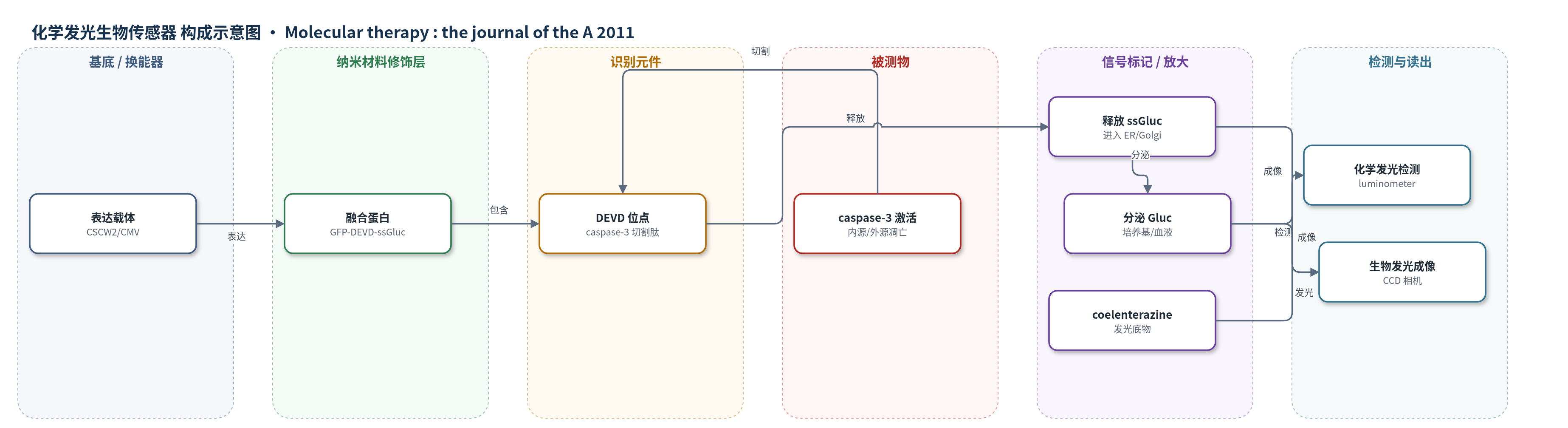
该传感器为基因编码融合蛋白 GFP-DEVD-ssGluc。未凋亡时,GFP 使含信号肽的 Gluc 滞留胞质,无法正确折叠,活性极低;凋亡诱导内源或外源通路后,caspase-3 被激活并特异性切割 DEVD 位点,释放 ssGluc。ssGluc 进入内质网、高尔基体和囊泡等经典分泌途径,完成折叠并分泌至条件培养基或血液。加入 coelenterazine 后,分泌的 Gluc 催化底物氧化产生光子,凋亡程度或 caspase-3 活性越高,分泌 Gluc 越多,化学发光/生物发光信号越强。体内注射 coelenterazine 后可用 CCD 相机成像。Gaussia 荧光素酶高灵敏度与分泌途径的活性开关构成信号放大。
检测灵敏度
原文未报告 LOD、线性范围、灵敏度斜率或相关系数;仅报告相对灵敏度:Gluc 比 Fluc 高 >2,000 倍,比分泌型碱性磷酸酶高 >20,000 倍。
效应效果
该传感器可特异性响应 caspase-3:ZVAD-fmk 抑制后 Gluc 释放消失;BFA 阻断分泌途径后胞外信号下降、胞内积累,证明经 ER/Golgi 分泌。可检测内源(阿霉素)与外源(TRAIL)凋亡,条件培养基信号随剂量增加,3.2 µmol/l 阿霉素使 Gluc 活性增加 >10 倍。裸鼠皮下肿瘤中,10 µg 阿霉素处理 8 h 后肿瘤 Gluc 信号约增加 5 倍;5 µl 血液检测在 8 h 达峰值,P<0.001。乳腺癌转移模型中 TRAIL 0.5 mg/kg 后 6 h 血液 Gluc 显著升高,48 h 回落。相比 Fluc 报告系统约 2–3 倍变化,本系统信号更强且可实时动力学监测,适合高通量筛选与临床前药效评价。
传感器的构成
- 表达载体/换能器:CSCW2 慢病毒载体(lenti-GFP-DEVD-ssGluc),CMV 启动子驱动融合蛋白在细胞内表达
- 识别元件:DEVD 肽(Asp-Glu-Val-Asp),作为 caspase-3 特异性切割位点,凋亡时被切割
- 信号报告蛋白:Gaussia 荧光素酶(Gluc)含信号肽 ssGluc,切割后进入分泌途径并分泌到胞外
- 胞质滞留/荧光标签:Ptilosarcus GFP(PtGFP/GFP),融合于 N 端使未切割融合蛋白滞留胞质且 Gluc 失活
- 发光底物:coelenterazine,与分泌的 Gluc 反应产生生物发光/化学发光信号
- 样品基质/读出介质:细胞条件培养基或小鼠血液,用于体外检测 Gluc 活性;体内成像用 CCD 相机
中文摘要
非侵入性检测与体内成像凋亡在癌症等多种疾病治疗开发中至关重要。作者将绿色荧光蛋白(GFP)融合到天然分泌型 Gaussia 荧光素酶(Gluc)N 端,中间以 caspase-3 切割肽 DEVD 连接,构建凋亡生物传感器。该融合蛋白以无活性形式滞留于胞质;凋亡发生时,激活的 caspase-3 切割 DEVD,释放含信号肽的 ssGluc。ssGluc 进入内质网、高尔基体和囊泡等分泌途径,正确折叠后从细胞分泌,可在培养条件培养基或活体动物血液中随时间检测。由于 Gluc 经经典分泌途径释放,其在分泌区室中的存在可用生物发光成像在体内可视化。该报告系统为凋亡成像与实时监测提供有价值工具,并适用于培养细胞和动物模型的高通量功能筛选。
英文摘要
Noninvasive detection and in vivo imaging of apoptosis plays a critical role in the development of therapeutics in many different fields including cancer. We have developed an apoptosis biosensor by fusing green fluorescent protein (GFP) to the N-terminus of the naturally secreted Gaussia luciferase separated by a caspase-3 cleavage peptide consisting of aspartic acid (D), glutamic acid (E), valine (V), and aspartic acid (D) or DEVD. We showed that this fusion is retained in the cytoplasm of cells in an inactive form. Upon apoptosis, the DEVD peptide is cleaved in response to caspase-3 activation, freeing ssGluc, which can now enter the secretory pathway where it is folded properly and is released from the cells and can be detected in the conditioned medium in culture or in blood of live animals ex vivo over time. Because Gluc is secreted from cells via conventional pathway through the endoplasmic reticulum (ER), Golgi and vesicles, we showed that the presence of Gluc in these compartments in response to apoptosis can be visualized in vivo using bioluminescence imaging. This reporter provides a valuable tool for imaging and real-time monitoring of apoptosis and is compatible with high-throughput functional screening application in cultured cells and animal models.