组成图示
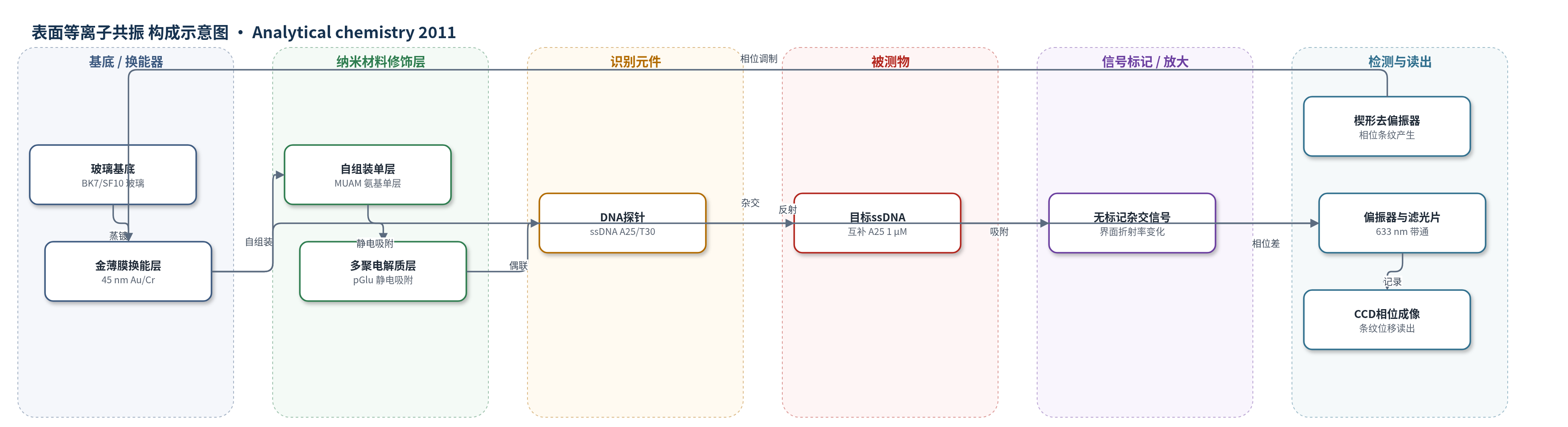
点击图片查看大图 · 依据论文自动绘制
传感器类型
表面等离子共振(SPR)生物传感器
检测对象
单链DNA(ssDNA,A25),样品基质为缓冲液(10 mM Na2HPO4、137 mM NaCl、2.7 mM KCl,pH 7.4)
检测原理
p 偏振光经楔形去偏振器形成空间周期相位调制,在 Kretschmann 棱镜/金膜界面激发表面等离子激元。金膜表面依次修饰 MUAM、pGlu 和 ssDNA 探针。目标 ssDNA 与互补探针杂交并吸附到金表面,改变界面局部折射率和表面覆盖度,使 p 偏振反射光的 Fresnel 相位 δp 发生变化。输出偏振器将相位变化转换为干涉条纹位置移动,CCD 记录条纹位移并计算相位差 Δφ。低浓度下表面覆盖度 Γ 与浓度 C 近似线性(Γ/Γmax=KadsC),因此 Δφ 随 ssDNA 浓度增加而增大。该体系不使用酶或纳米颗粒放大,主要依靠相位检测提高灵敏度。
检测灵敏度
LOD: 50 pM (Clod = 0.001/(2 × 10^7));最小可检测相位差: Δφ = 0.03°;DNA 单层响应: Δφ = 28.80 ± 0.03°;可检测覆盖: 0.1% of a DNA monolayer;MUAM 单层响应: Δφ = 182.0 ± 0.03°;可检测覆盖: ~0.02% of a MUAM monolayer
效应效果
该 SPR-PI 微阵列在 1 μM 目标 ssDNA 下实现实时杂交吸附监测,A25 目标仅与 T30 探针线产生相位差,A25 对照线无响应,显示序列特异性。完整 DNA 单层吸附给出 Δφ = 28.80 ± 0.03°,最小可检测相位差 0.03°,对应约 0.1% 单层覆盖和 50 pM 浓度检出限;传统 SPRI 对同一体系约 5 nM,因此灵敏度提高约 100 倍。作者认为其动态范围与强度型 SPR 相当,可用于高通量、原位、多通道生物亲和相互作用监测。
传感器的构成
- 基底/换能器:BK7 或 SF10 玻璃片、1 nm Cr 粘附层和 45 nm Au 薄膜,提供 SPR 换能界面
- 图案化金条纹:SF10 玻璃上 1 mm 宽、1.5 mm 周期 Au 条纹,形成线性微阵列传感区
- 自组装单层:11-巯基十二胺(MUAM)自组装单层,提供氨基界面并用于光刻图案化
- 多聚电解质修饰层:聚-L-谷氨酸钠盐(pGlu)静电吸附,连接 DNA 探针并调节界面
- DNA识别探针:氨基修饰 ssDNA(A25 或 T30)经 EDC/NHSS 偶联到 pGlu/MUAM,用于杂交捕获互补 ssDNA
- 信号标记:无外源标记,目标 ssDNA 直接改变金表面折射率并产生相位信号
- 光学读出:钨灯白光光源、楔形去偏振器、线性偏振器、633 nm 带通滤光片和 CCD,将相位差转换为条纹位移
中文摘要
本文报道将表面等离子共振相位成像(SPR-PI)用于线性微阵列格式,以实时测量表面生物亲和吸附过程。SPR-PI 在 ATR Kretschmann 构型中测量 p 偏振光在 SPR 角入射到金薄膜后反射光的相位偏移,并通过偏振器—石英楔去偏振器组合在界面形成干涉条纹图像。当生物分子吸附到金薄膜时,条纹位置发生移动。利用与干涉条纹垂直的 500 μm 生物传感元件线,可同时实时监测多个生物亲和吸附过程。作者首先通过光刻制备 10 条 11-巯基十二胺(MUAM)自组装单层,验证多通道相位差测量;1.8 nm MUAM 单层产生 Δφ = 182.08 ± 0.03°,与 Fresnel 方程和 Jones 矩阵计算一致。随后在六组分 DNA 线微阵列上检测单链 DNA(ssDNA)杂交吸附,完整 DNA 单层产生 Δφ = 28.80 ± 0.03°,并实现实时监测。结果表明 SPR-PI 的检测限有望比传统强度型 SPR 成像低约 100 倍。
英文摘要
The optical technique of surface plasmon resonance phase imaging (SPR-PI) is implemented in a linear microarray format for real-time measurements of surface bioaffinity adsorption processes. SPR-PI measures the phase shift of p-polarized light incident at the SPR angle reflected from a gold thin film in an ATR Kretschmann geometry by creating an interference fringe image on the interface with a polarizer-quartz wedge depolarizer combination. The position of the fringe pattern in this image changes upon the adsorption of biomolecules to the gold thin film. By using a linear array of 500 μm biosensor element lines that are perpendicular to the interference fringe image, multiple bioaffinity adsorption measurements can be performed in real time. Two experiments were performed to characterize the sensitivity of the SPR-PI measurement technique: First, a ten line pattern of a self-assembled monolayer of 11-mercaptoundecamine (MUAM) was created via photopatterning to verify that multiple phase shifts could be measured simultaneously. A phase shift difference (Δφ) of Δφ = 182.08 ± 0.03° was observed for the 1.8 nm MUAM monolayer; this value agrees with the phase shift difference calculated from a combination of Fresnel equations and Jones matrices for the depolarizer. In a second demonstration experiment, the feasibility of SPR-PI for in situ bioaffinity adsorption measurements was confirmed by detecting the hybridization and adsorption of single stranded DNA (ssDNA) onto a six-component DNA line microarray patterned monolayer. Adsorption of a full DNA monolayer produced a phase shift difference of Δφ = 28.80 ± 0.03° at the SPR angle of incidence and the adsorption of the ssDNA was monitored in real time with the SPR-PI. These initial results suggest that SPR-PI should have a detection limit roughly 100 times lower than traditional intensity-based SPR imaging measurements.