组成图示
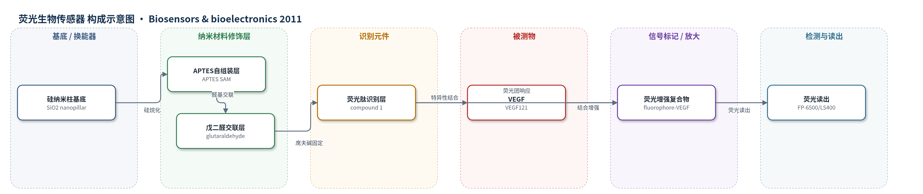
点击图片查看大图 · 依据论文自动绘制
传感器类型
荧光生物传感器
检测对象
血管内皮生长因子(vascular endothelial growth factor, VEGF,重组人VEGF121);样品基质:大鼠血清(rat serum)
检测原理
该传感器以二氧化硅纳米柱为基底,通过APTES自组装单层和戊二醛交联层固定荧光肽compound 1。荧光肽同时包含VEGF结合肽段和荧光团,未结合VEGF时荧光较弱;当VEGF与肽段结合后,荧光团进入VEGF疏水残基形成的微环境,受溶剂极性和去溶剂化影响,激发态与基态能量差改变,荧光量子产率显著增加,产生约525 nm绿色荧光增强。纳米柱结构将表面积增大至平面基底约6倍,使固定肽量增加,从而提高信号。荧光强度随VEGF浓度增加而线性增强,结合亲和力Kd为6.0×10⁻⁹ M,无需酶催化或核酸放大,信号放大主要来自纳米柱表面积增强和荧光团环境响应。
检测灵敏度
LOD: below 1.0 ng/mL;线性范围: 0–0.05 μg/mL(0–50 ng/mL);R^2 > 0.996;Kd: 6.0 × 10⁻⁹ M
效应效果
该传感器在大鼠血清中检测VEGF,具有较高选择性:0–0.5 μg/mL牛血清白蛋白(BSA)以及转铁蛋白、IgG均未引起荧光增强。三次重复校准的变异系数(CV)在3.8%以内,表明重现性良好。4 ℃冷冻保存约1个月后,对VEGF的响应仍保持初始值的约95%,稳定性较好。与相同材料制备的平面基底相比,纳米柱基底在0.5 μg/mL VEGF下的荧光强度约为平面的5倍;纳米柱可检测低于1.0 ng/mL的VEGF,而平面基底在5.0 ng/mL时未检出。作者认为该荧光纳米柱系统可便捷、快速、特异地监测血管生成生物标志物VEGF,适用于医院、家庭等场景。
传感器的构成
- 基底:硅晶圆经电子束光刻(EBL)与电感耦合等离子体反应离子刻蚀(ICP-RIE)形成硅纳米柱阵列,再热氧化为二氧化硅纳米柱(silica nanopillar)基底,提供约6倍表面积。
- 硅烷偶联层:3-aminopropyltrimethoxysilane(APTES)在甲苯中形成氨基自组装单层(SAM),用于连接纳米柱表面与后续交联层。
- 交联层:戊二醛(glutaraldehyde, GA)与SAM氨基反应生成醛基,用于共价固定荧光肽。
- 识别元件:compound 1中的VEGF结合肽段(GPGSGRGWVEICAADDYGRGPGSK)特异性识别VEGF。
- 信号标记物:compound 1中的荧光团(fluorophore)在结合VEGF后进入疏水微环境,荧光增强。
中文摘要
本研究建立了一种结合纳米制造技术的高灵敏、高选择性蛋白检测技术。作者采用电子束光刻和深反应离子刻蚀制备了间距200 nm、高度1000 nm的二氧化硅纳米柱芯片,并将对血管内皮生长因子(VEGF)具有高亲和力的荧光肽通过3-氨基丙基三乙氧基硅烷和戊二醛自组装单层固定在纳米柱表面。在荧光分光光度计和荧光扫描图像分析下,纳米柱上荧光肽的荧光强度随VEGF浓度增加而增强;非线性最小二乘拟合得到的解离常数Kd为6.0×10⁻⁹ M,有助于高灵敏检测VEGF。与平面基底相比,密集且较高的纳米柱阵列增大了虚拟蛋白结合面积,使结合VEGF后的荧光强度更高。重现性测试和寿命测量表明,该荧光试剂可作为检测VEGF的有用生物传感器。结果表明,荧光试剂与纳米柱基底的组合可作为一种便捷方法用于VEGF检测。
英文摘要
We have established a highly sensitive and selective protein detection technology in combination with the nanofabrication technique. A silica nanopillar chip with a 200-nm pitch and 1000-nm height pillar substrate was fabricated by electron beam lithography and deep reactive ion etching method. Fluorescent peptides, with high affinity towards vascular endothelial growth factor (VEGF), were immobilized on nanopillar chip via a self-assembled monolayer made from 3-aminopropyltrimethoxysilane and glutaraldehyde under optimal conditions. The fluorescence intensity of the fluorescent peptide on the nanopillar substrate increased with increasing VEGF concentrations, as determined by a fluorescence spectrophotometer and fluorescent scanning image analysis. The dissociation constant (K(d) value) calculated by the non-linear least square curve fitting method was 6.0 × 10(-9)M, which contributed to the highly sensitive detection of VEGF. The fluorescence intensity of the fluorescent reagent on the nanopillar substrate upon binding to VEGF was higher than that obtained using the flat substrate because the dense and tall nanopillar array increased the virtual protein binding area. The reproducibility tests and lifetime measurement indicate the fluorescent reagent to be a useful biosensor for the detection of VEGF in this system. These experimental results clearly showed that the combination of a fluorescent reagent and a nanopillar substrate may be widely applicable as a convenient method for the detection of VEGF.