组成图示
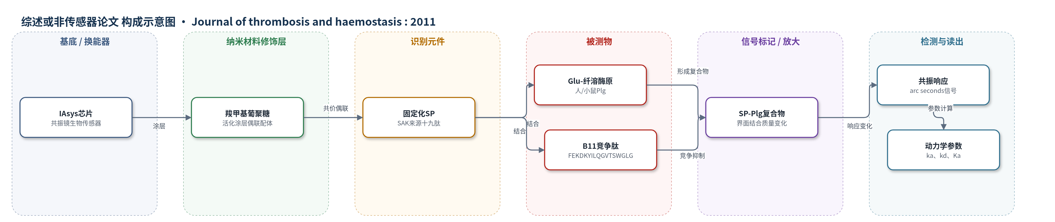
点击图片查看大图 · 依据论文自动绘制
传感器类型
综述或非传感器论文
检测对象
纤溶酶原(plasminogen, Glu-plasminogen/Lys-plasminogen)、B11肽(FEKDKYILQGVTSWGLG)、组织型纤溶酶原激活物(t-PA);样品基质:磷酸盐缓冲液、人/小鼠血浆或血栓模型血液
检测原理
SP 与 Glu-纤溶酶原 B 区结合,关键残基 Asp750 与 SP 中 Lys14 可能通过静电作用识别,诱导纤溶酶原局部构象变化,使 t-PA 更易接近切割位点,降低 Km 并提高 kcat/Km。在 IAsys 共振镜生物传感器中,SP 或纤溶酶原共价固定于羧甲基葡聚糖涂层芯片,溶液相结合物结合后改变界面质量/折射率,引起共振响应(arc seconds)变化,实时获得结合与解离动力学。比色法中,t-PA 将纤溶酶原转化为纤溶酶,纤溶酶水解 S-2251 释放 p-硝基苯胺,405 nm 吸光度随纤溶酶生成量增加,从而反映激活效率。
检测灵敏度
原文未报告 LOD、线性范围、灵敏度斜率或相关系数。
效应效果
SP 选择性增强 t-PA 对 Glu-纤溶酶原激活,不增强 Lys-纤溶酶原激活,也不改变纤溶酶活性;37°C 缓冲液 7 天保留 >80% 活性,与 plasmin-Sepharose 或 t-PA-Sepharose 孵育 1 h 后活性完全保留。B11 肽抑制 SP 介导激活,D750A/D750K/DD750 突变显著降低抑制活性,提示 Asp750 关键。小鼠颈动脉血栓模型中,SP 剂量依赖促进再通:62.5、125、250 μg/kg 再通时间分别为 2.38±1.11、9.25±2.12、13.45±6.14 min,对照 0.67±0.55 min;t-PA 基因缺失小鼠无再通,PAI-1 缺失小鼠再通更早。SP 不引起血浆纤维蛋白原降解,作者认为其可作为 t-PA 替代或增效剂。
传感器的构成
- 基底/换能器:IAsys 共振镜生物传感器芯片,提供光学共振检测界面
- 修饰层:活化羧甲基葡聚糖涂层(carboxymethyl dextran-coated cuvette),用于共价偶联蛋白或肽
- 识别元件:固定化 SP 或 Glu-plasminogen/t-PA/u-PA,作为结合分析配体
- 目标物:溶液相 Glu-plasminogen、B11 肽或内皮细胞,用于检测结合
- 信号读出:共振响应(arc seconds)随界面结合质量/折射率变化而改变
中文摘要
背景:此前发现一种源自葡萄球菌激酶(SAK)的合成十九肽 SP(GPYLMVNVTGVDGKGNELL)可增强 SAK/纤溶酶复合物对纤溶酶原的激活。目的:鉴定 SP 在纤溶酶原上的结合位点,并阐明 SP 对组织型纤溶酶原激活物(t-PA)激活纤溶酶原的影响。方法:采用比色底物法和还原条件 SDS-PAGE 检测纤溶酶原激活;用 IAsys 生物传感器分析 SP 与纤溶酶原 B 区来源肽段的结合;用圆二色光谱分析 SP 诱导的纤溶酶原结构变化;在小鼠血栓模型中评价溶栓效果。结果:SP 以浓度依赖方式增强 t-PA 对 Glu-纤溶酶原的激活,催化效率(kcat/Km)提高 11.4 倍;位于纤溶酶原 B 区近 C 端的合成肽 FEKDKYILQGVTSWGLG 显著抑制 SP 与纤溶酶原结合;SP 与 Glu-纤溶酶原复合物的近紫外 CD 谱与 Glu-纤溶酶原明显不同;SP 在小鼠血栓模型中剂量依赖地促进早期再通,但在 t-PA 基因缺失小鼠中无效。结论:SP 结合纤溶酶原 B 区并促进 t-PA 激活纤溶酶原,从而诱导有效溶栓。
英文摘要
BACKGROUND: A synthetic nonadecapeptide (SP; GPYLMVNVTGVDGKGNELL) previously enhanced the activation of plasminogen by the SAK/plasmin complex.
OBJECTIVES: To identify the binding site for SP on plasminogen and elucidate the effects of SP on plasminogen activation by the tissue-type plasminogen activator (t-PA).
METHODS: The effects of SP on plasminogen activation were estimated using a chromogenic substrate and from the cleavage of plasmin on SDS-PAGE under reduced conditions. The binding to SP of various peptides derived from the amino acid sequence of plasminogen was analyzed with an IAsys biosensor. The SP-mediated structural change to plasminogen was analyzed by circular dichroism (CD) spectroscopy. The thrombolytic effects of SP were examined using a mouse model of thrombosis.
RESULTS: SP enhanced the activation of plasminogen by t-PA. The catalytic efficiency (k(cat)/K(m)) of Glu-plasminogen activation by t-PA was 11.4-fold higher in the presence than absence of SP. The binding of SP to plasminogen was greatly inhibited by a synthetic peptide, FEKDKYILQGVTSWGLG, located close to the C-terminal of the plasminogen B region. Near-ultraviolet CD spectra of the complex between SP and Glu-plasminogen significantly differed from those of Glu-plasminogen. When SP was administered in a mouse model of thrombosis, early recanalization was observed in a dose-dependent manner. However, SP did not cause recanalization in t-PA gene-deficient mice.
CONCLUSIONS: SP bound to the B region and promoted the activation of plasminogen by t-PA, and then induced effective thrombolysis.