组成图示
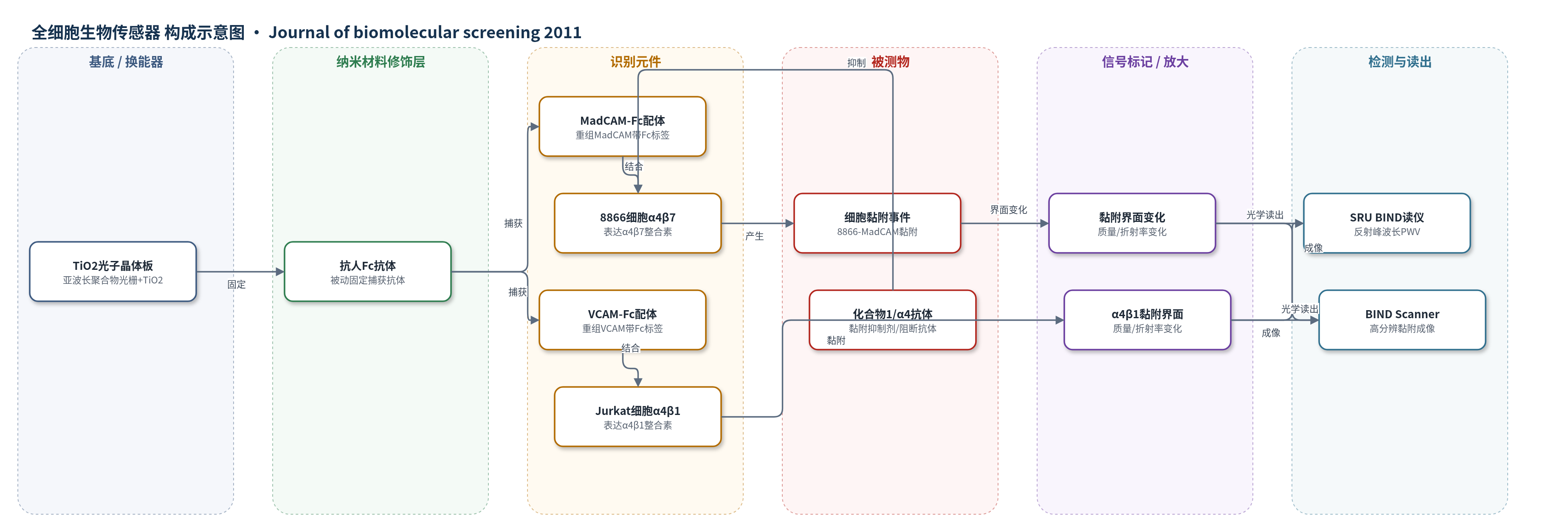
点击图片查看大图 · 依据论文自动绘制
传感器类型
全细胞生物传感器
检测对象
α4β7介导的细胞黏附(α4β7-mediated cell adhesion,8866细胞至MadCAM)、α4β1介导的细胞黏附(α4β1-mediated cell adhesion,Jurkat细胞至VCAM);样品基质为含二价阳离子的细胞悬液/缓冲液(HEPES-NaCl-CaCl2-MgCl2)。
检测原理
SRU BIND传感器以TiO2涂覆的亚波长聚合物光栅为光学换能器,共振反射单一波长。抗人Fc抗体被动固定于TiO2表面,定向捕获带Fc标签的重组MadCAM或VCAM。8866细胞表面α4β7整合素与MadCAM特异性结合,该过程依赖Mg2+/Ca2+;细胞黏附在传感器表面后增加界面质量负载并改变局部折射率,使反射峰波长值(PWV)发生偏移。无标记条件下,黏附细胞数量与黏附强度越大,PWV偏移越大;化合物1或α4阻断抗体与细胞孵育后降低整合素亲和力或阻断结合,使黏附减少,PWV偏移降低。Reader以30 s间隔记录PWV动力学,Scanner可成像单细胞黏附强度。
检测灵敏度
相关系数: 0.86(手动法与SRU BIND法IC50比较);相关系数: 0.87(两次SRU BIND测试)
效应效果
该无标记平台选择性良好:α4阻断抗体21/6(15 mg/mL)完全阻断8866细胞黏附MadCAM,β7抗体亦阻断;无MadCAM或BSA封闭孔仅产生小PWV偏移。黏附严格依赖二价阳离子,Mg2+促进强于Ca2+。与手动荧光法相比,标准化合物1的IC50分别为238 nM和430 nM;一系列化合物IC50相关系数0.86,Z′分别为0.61和0.56;两次独立测试相关系数0.87。BIND Scanner显示化合物1剂量依赖降低成像细胞数和黏附强度,IC50为340 nM,且明场计数表明沉降细胞数不变,说明主要成像黏附细胞。作者认为该平台适合高通量筛选整合素拮抗剂,可用于炎症疾病和癌症药物发现。
传感器的构成
- 基底/换能器:TiO2涂层384孔板(SRU BIND板),亚波长聚合物光栅涂覆高折射率TiO2,光学换能,反射峰波长随表面生物材料吸附变化。
- 捕获抗体层:抗人Fc抗体(Sigma cat. I-6260),被动固定于TiO2表面,用于定向捕获带Fc标签的MadCAM/VCAM。
- 配体识别层:重组MadCAM-Fc(0.15 mg/mL)或VCAM-Fc(0.1 mg/mL),过夜孵育,作为细胞表面整合素识别配体。
- 封闭层:4%热灭活BSA(buffer 1中)封闭非特异结合位点;BSA封闭板用于定义被动沉降背景。
- 全细胞识别元件:8866人B细胞(内源表达α4β7整合素)或Jurkat细胞(表达α4β1),作为全细胞识别元件。
- 信号标记/放大:无标记,细胞黏附本身改变表面折射率/质量负载,产生PWV偏移;无荧光/酶标记。
- 读出系统:SRU BIND Reader宽谱光源、光纤、光谱仪,测量反射峰波长值PWV;BIND Scanner成像。
中文摘要
传统细胞黏附实验通常采用手动工作站和荧光读出。本文报道了一种基于SRU BIND平台的无标记均相检测方法,用于筛选α4β7整合素介导的细胞黏附至其配体黏膜地址素细胞黏附分子(MadCAM)的抑制剂。该生物传感器为光学型,核心为亚波长聚合物光栅,并涂覆高折射率TiO2材料;细胞特异性黏附引起反射峰波长值(PWV)偏移。实验使用标准化合物和α4阻断抗体验证,与手动荧光法在一系列不同效力化合物上高度一致。细胞黏附严格依赖二价阳离子,Mg2+促进黏附强于Ca2+。该无标记均相格式变异低,Z′为0.6。除检测表达α4β7的8866细胞黏附MadCAM外,还证明该平台可检测表达α4β1的Jurkat细胞黏附血管细胞黏附分子(VCAM),表明其适用于高通量筛选其他整合素介导的细胞黏附事件。
英文摘要
Traditionally, cell adhesion assays are performed in a manual workstation format using fluorescence-based readouts. Herein, the authors describe a label-free homogeneous assay to identify inhibitors of α4β7 integrin-mediated cell adhesion to its ligand, the mucosal addressin cell adhesion molecule (MadCAM), using the SRU BIND platform. The biosensor is optically based and comprises a subwavelength polymer grating. The assay was validated using standard compounds and an α4 blocking antibody and correlated very closely with the manual assay format when running a battery of test compounds of varying potencies. Cell adhesion was strictly dependent on the presence of divalent cations where Mg(2+) was greater than Ca(2+) at promoting cell adhesion. This homogeneous and label-free format exhibited low variability with a calculated Z' of 0.6. In addition to measuring α4β7-mediated 8866 cell adhesion to MadCAM, the authors also demonstrate that this platform can measure adhesion of Jurkat cells expressing α4β1 to the vascular cell adhesion molecule. Thus, the SRU BIND platform is widely applicable to measuring cell adhesion events mediated by other integrins binding to their receptors in an assay format that is amenable to high-throughput screening.