组成图示
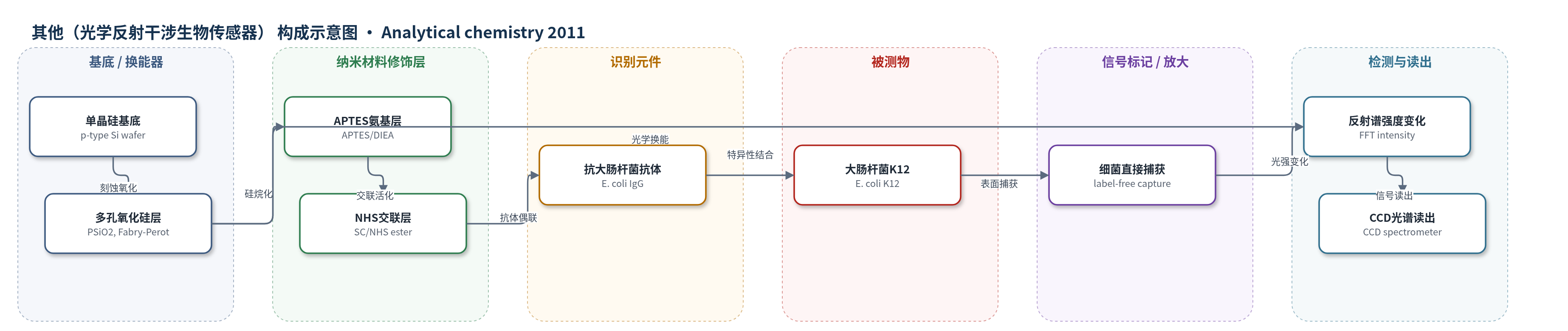
点击图片查看大图 · 依据论文自动绘制
传感器类型
其他(光学反射干涉生物传感器)
检测对象
大肠杆菌K12(Escherichia coli K12);样品基质:细菌悬液(LB培养后)
检测原理
该传感器以氧化多孔硅(PSiO2)薄膜作为Fabry–Perot光学干涉换能器。PSiO2表面经APTES和SC交联剂共价固定抗大肠杆菌IgG,形成识别层。当样品中的E. coli K12与表面抗体特异性结合时,细菌被直接捕获在多孔表面;由于细菌尺寸(约0.8–2 μm)大于孔径(60–100 nm),结合主要发生在表面而非孔内。细菌附着改变薄膜表面/界面光学性质,使反射谱FFT峰强度发生可预测下降,且强度变化与细菌浓度成正比。该过程无需荧光、酶或纳米标记物,也不依赖酶催化放大,主要依靠多孔硅高比表面积和抗体特异性捕获实现信号增强,可在数分钟内实时读出。
检测灵敏度
LOD: 10^4 cells/mL;线性范围: 10^3–10^5 cells/mL
效应效果
实验比较了特异性共价固定与物理吸附固定抗体的PSiO2传感器,在10^3–10^5 cells/mL范围内,特异性固定传感器均产生更大反射谱强度变化,未修饰PSiO2对照变化不显著,说明响应来自抗体介导的直接细胞捕获。荧光标记对照显示固定抗体保留免疫活性且无交叉反应。当前检测限为10^4 cells/mL,响应时间为数分钟;10^4 cells/mL时FFT强度下降3.4±0.5%。作者指出SPR细菌传感器检测限通常为10^2–10^6 cells/mL,本平台响应时间相当且结构更简单。论文未报告RSD、实际样品回收率或长期稳定性,但认为可推广至多种微生物检测。
传感器的构成
- 基底/换能器:高掺杂p型单晶硅片(p-type Si wafer),经HF/乙醇电化学刻蚀并800℃热氧化,形成多孔氧化硅光学换能基底
- 多孔氧化硅层:氧化多孔硅(PSiO2),厚度约7880±60 nm、孔隙率约80%、孔径60–100 nm,构成Fabry–Perot薄膜干涉光学响应
- 氨基硅烷修饰层:3-氨基丙基三乙氧基硅烷(APTES)在二异丙基乙胺(DIEA)催化下接枝到PSiO2表面,引入氨基
- 交联活化层:双(N-琥珀酰亚胺基)碳酸酯(SC)与APTES氨基反应形成NHS活性酯,用于抗体共价偶联
- 识别元件:抗大肠杆菌单克隆抗体/免疫球蛋白G(E. coli IgG)通过氨基与NHS酯共价固定,特异性捕获细菌
- 清洗/非特异去除:PBS缓冲液与1 M NaCl洗涤,去除未结合抗体及非特异吸附
- 信号标记物:无标记(label-free),细菌本身作为光学信号源,不添加荧光、酶或纳米标记物
中文摘要
本文提出一种基于纳米结构氧化多孔硅(PSiO2)的光学无标记细菌检测生物传感平台,以大肠杆菌K12为模型体系。该传感器无需细胞裂解等样品前处理,即可通过表面特异性抗体直接捕获目标细菌。PSiO2纳米结构作为光学换能元件,其反射谱呈现薄膜干涉特有的Fabry–Perot条纹,可在数分钟内实时观察细菌附着。多孔表面经共价偶联特异性单克隆抗体(IgG)形成识别层。作者利用衰减全反射傅里叶变换红外光谱、荧光标记实验和折射干涉傅里叶变换光谱证实抗体成功固定,并保留免疫活性与特异性。目标细菌与表面抗体结合后发生直接细胞捕获,引起薄膜光学干涉谱可预测变化。初步实验表明该平台可用于低浓度细菌检测,当前对E. coli K12的检测限为10^4 cells/mL,响应时间为数分钟。
英文摘要
An optical label-free biosensing platform for bacteria detection ( Escherichia coli K12 as a model system) based on nanostructured oxidized porous silicon (PSiO(2)) is introduced. The biosensor is designed to directly capture the target bacteria cells on its surface with no prior sample processing (such as cell lysis). The optical reflectivity spectrum of the PSiO(2) nanostructure displays Fabry-Pérot fringes characteristic of thin-film interference, enabling direct, real-time observation of bacteria attachment within minutes. The PSiO(2) optical nanostructure is synthesized and used as the optical transducer element. The porous surface is conjugated with specific monoclonal antibodies (immunoglobulin G's) to provide the active component of the biosensor. The immobilization of the antibodies onto the biosensor system is confirmed by attenuated total reflectance Fourier transform infrared spectroscopy, fluorescent labeling experiments, and refractive interferometric Fourier transform spectroscopy. We show that the immobilized antibodies maintain their immunoactivity and specificity when attached to the sensor surface. Exposure of these nanostructures to the target bacteria results in "direct cell capture" onto the biosensor surface. These specific binding events induce predictable changes in the thin-film optical interference spectrum of the biosensor. Our preliminary studies demonstrate the applicability of these biosensors for the detection of low bacterial concentrations. The current detection limit of E. coli K12 bacteria is 10(4) cells/mL within several minutes.