组成图示
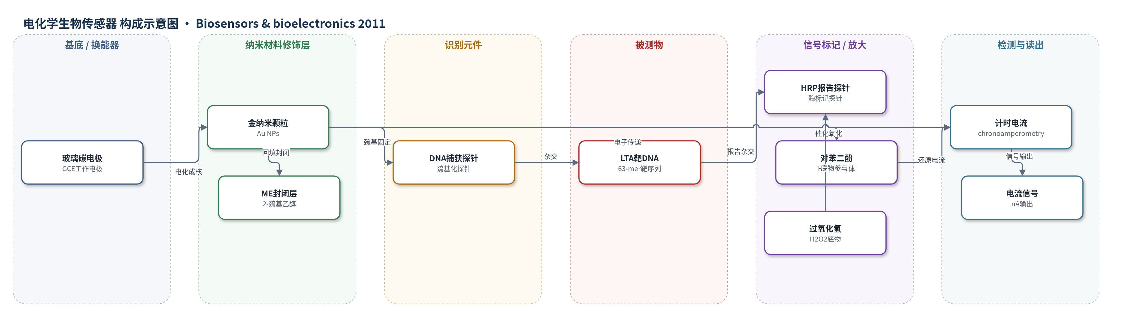
点击图片查看大图 · 依据论文自动绘制
传感器类型
电化学生物传感器
检测对象
淋巴毒素-α基因靶序列(LTA target DNA / LTA amplicon);样品基质:PBS-600杂交缓冲液(10 mM PBS、600 mM NaCl、pH 7.4)中的合成寡核苷酸溶液
检测原理
传感器以GCE为基底,通过KAuCl4电化成核形成Au NPs纳米结构,再用巯基化DNA捕获探针和ME构建混合自组装单分子层。Au NPs提供纳米级固定域,控制探针间距与取向,减少杂交位阻并增强电子传递。LTA靶DNA与捕获探针杂交后,HRP标记报告探针结合形成三明治复合物。检测时加入HQ和H2O2,HRP催化H2O2氧化HQ生成BQ,HQ在-0.1 V下于电极还原,产生与靶浓度成正比的计时电流。纳米金表面通过提高探针密度、催化活性和电子中继效应实现信号放大。
检测灵敏度
LOD: 0.53 nM;动态检测范围: 0.53 nM–25 nM;灵敏度: 44.89 nA × nM−1;平面金电极 LOD: 5.22 nM,灵敏度: 13.67 ± 1.74 nA × nM−1
效应效果
三轮沉积电极最佳,LOD 0.53 nM,灵敏度44.89 nA×nM−1,动态范围0.53–25 nM;平面金电极LOD 5.22 nM,灵敏度13.67±1.74 nA×nM−1,检出限降低约一个数量级。非互补靶10、50、100 nM信号20.33±0.56 nA,与空白相当,无交叉反应。SEM显示三轮后Au NPs密度约23个/µm²,粒径80.86±4.16 nm。第四轮未提升性能,提示覆盖过高导致位阻。电极90°C热水洗3 min可再生。未报告实际样品回收率、RSD或与ELISA对比,作者认为适用于DNA检测及多路复用。
传感器的构成
- 基底/换能器电极:玻璃碳电极(GCE),抛光并电化学活化,作为工作电极与电子传导基底
- 纳米材料修饰层:金纳米颗粒(Au NPs),由KAuCl4电化成核形成随机纳米金域,提高探针固定密度与电子/催化性能
- 识别元件:5′-巯基化DNA捕获探针(thiolated DNA capture probe),通过Au-S键固定于Au NPs,与LTA靶序列杂交
- 封闭/绝缘层:2-巯基乙醇(ME),回填未覆盖金表面,抑制非特异吸附并绝缘Au NPs以支持多轮成核
- 信号标记物:HRP标记报告探针(HRP-conjugated reporter probe),与靶-捕获双链杂交,提供酶催化信号
- 电子供体/底物:对苯二酚(HQ)与过氧化氢(H2O2),HRP催化HQ氧化为苯醌(BQ),HQ在电极还原产生电流
中文摘要
生物传感器读出信号可通过优化换能器表面设计,使固定识别元件与目标分析物实现最佳相互作用而增强。基因传感器中,固定DNA捕获探针的间距与取向需精确控制,以最大化表面杂交并结合信号。本文提出一种基于玻璃碳电极(GCE)表面纳米结构化的新方法,用于开发高灵敏电化学生物传感器。通过电化学成核在GCE上依次沉积金纳米颗粒,形成随机分布的致密金纳米域;通过交替进行短程成核和巯基化DNA探针绝缘,可提高成核颗粒数密度并防止聚集。以淋巴毒素-α(LTA)基因检测为模型,采用HRP标记报告探针的三明治格式进行安培检测。三轮沉积电极灵敏度最高,为44.89 nA×nM−1,动态检测范围为0.53–25 nM,比平面金电极低一个数量级。该纳米结构表面可用于DNA生物传感器及其他高灵敏检测。
英文摘要
Biosensor read out signals can be enhanced by carefully designing the transducer surfaces to achieve an optimal interaction between the recognition elements immobilised and the targeted analyte. This is particularly evident in the case of genosensors, where spacing and orientation of immobilised DNA capture probes need to be controlled to maximise subsequent surface hybridisation with the target sequence and achieve high binding signals. Addressing this goal, we present a novel approach based on the surface nanostructuring of glassy carbon electrodes (GCEs) towards the development of highly sensitive electrochemical genosensors. Gold nanoparticles were sequentially electrochemically nucleated on glassy carbon electrodes to form dense arrays of randomly distributed gold nanodomains. The number density of the electronucleated nanoparticles could be increased by repeatedly alternating between a short electronucleation step and the subsequent insulation of the nucleated nanoparticles with thiolated DNA probes. This approach allowed for the creation of highly structured surfaces whilst preventing aggregation of nanoparticles. The performances of planar gold electrodes and that of the nanopatterned surfaces prepared following several rounds of deposition were compared for the amperometric detection of DNA. Three rounds of deposition exhibited the highest sensitivity (44.89 nA × nM(-1)), with a dynamic detection range spanning from 0.53 nM to 25 nM of the targeted sequence, i.e. one order of magnitude lower than that obtained for the planar gold electrodes. The use of the nanostructured surface we report here may find application not only in DNA biosensors but also for any sensing application requiring highly sensitive measurements.