组成图示
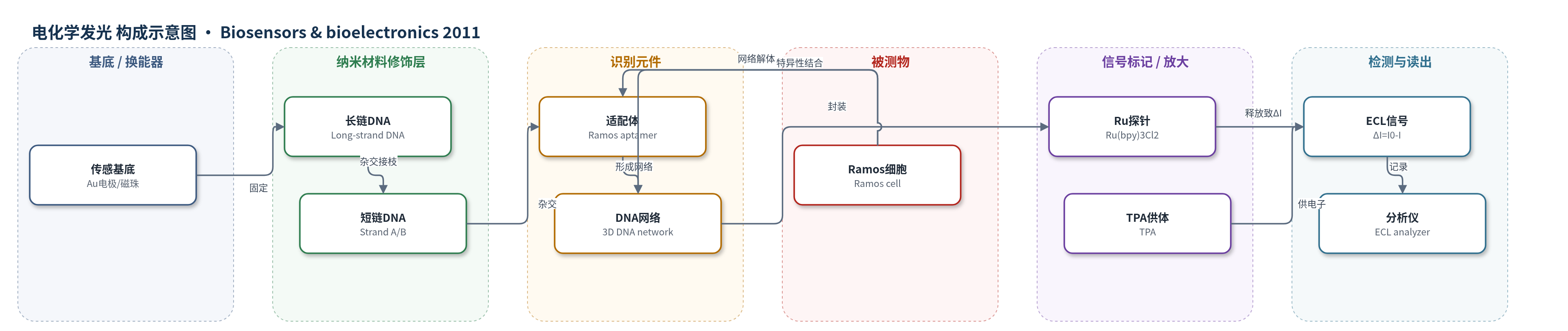
点击图片查看大图 · 依据论文自动绘制
传感器类型
电化学发光(ECL)生物传感器
检测对象
Ramos 癌细胞(Ramos cell);样品基质为 RPMI 1640 细胞培养基/PBS 重悬的细胞悬液,另用 CEM、K562 细胞作对照
检测原理
传感器以金电极或磁珠为界面,长链 DNA 固定后与短链 A/B 杂交形成 L-A/L-B。加入 Ramos 细胞适配体后,适配体同时与 L-A、L-B 杂交,使四种 DNA 形成三维网络,并将 Ru(bpy)3Cl2 物理封装于网络中。未加细胞时,电极表面保留大量 Ru,在 100 mM PBS 和 TPA 存在下,Ru(bpy)3^2+ 在约 1.14 V 发生电化学氧化并与 TPA 氧化产物反应产生 ECL,信号为 I0。加入 Ramos 细胞后,适配体与细胞表面靶标特异性结合,竞争性地从 DNA 网络中解离,导致网络解体并释放 Ru;电极表面 Ru 减少,ECL 峰高下降,ΔI=I0-I 随细胞浓度增加而增大。由于一个细胞可结合多个适配体并触发大量 Ru 释放,实现信号放大。
检测灵敏度
LOD: 58 cells/mL;线性范围: 100–1000 cells/mL;斜率: 1.02;方程: ΔI = 1.02 C + 114.77;R^2 = 0.9994;指数范围: 100–30,000 cells/mL,回归系数 0.9991
效应效果
该传感器对 Ramos 细胞具有良好选择性:CEM 和 K562 细胞几乎无响应,等量 Ramos 与 CEM 或 K562 混合样品的 ΔI 与单独 Ramos 相近。稳定性方面,4 ℃保存 24 h 后 I0 保留 98.1%,1000 cells/mL 的 ΔI 保留 97.4%;室温 PBS 中浸泡 6 h 后 ECL 仅下降 2.2%。重复性方面,1000 cells/mL 七次测量 RSD 为 3.2%。与文献方法相比,ECL 模式 LOD 58 cells/mL,优于 Medley 等 300 cells/mL、Smith 等 1250 cells/mL、Du 等 1.0×10^4 cells/mL,也优于 Ding 等荧光法 100 cells/mL;总检测时间约 1 h,操作简便、成本低,作者认为可用于便携式低成本癌症检测。
传感器的构成
- 基底/换能器:金电极(Au electrode)或羧基磁珠(Carboxyl modified MBs),作为传感界面
- 长链DNA修饰层:5′-巯基/氨基长链DNA(Long-strand DNA,含五个CCTACCTGCATGA重复单元),固定于Au或MBs
- 短链DNA接枝层:短链DNA A(Strand A)和短链DNA B(Strand B),与长链杂交形成L-A和L-B
- 识别元件:Ramos细胞适配体(Aptamer),与L-A/L-B杂交形成三维网络并特异性结合Ramos细胞
- 信号标记物:Ru(bpy)3Cl2(Ru)被DNA三维网络物理封装,作为ECL信号探针;文中另以FCA或luminol验证普适性
- 封闭剂:6-巯基-1-己醇(MCH),封闭未覆盖金表面,减少非特异性吸附
- 电子供体:三丙胺(TPA),在ECL测量中与Ru(bpy)3^2+反应产生发光
中文摘要
本文报道了一种用于检测 Ramos 癌细胞的无标记传感技术,其核心是以 DNA 三维网络封装三(2,2'-联吡啶)二氯化钌(II)(Ru(bpy)3Cl2,简称 Ru)作为信号探针。首先以金电极或羧基磁珠为传感界面,修饰含五个重复单元的长链 DNA;随后通过杂交分别将短链 DNA A 和 B 接枝到长链上,形成 L-A 和 L-B。加入 Ramos 细胞适配体后,适配体与 L-A、L-B 杂交,使四种 DNA 最终形成三维网络结构,同时 Ru 被物理封装于网络中。当 Ramos 细胞加入时,细胞与适配体特异性结合,导致 DNA 网络解体并释放 Ru,使电化学发光(ECL)信号下降。为验证方法的普适性,作者还分别以 FCA(ferrocenecarboxylic acid)和鲁米诺(luminol)作为信号探针,构建计时库仑(CC)和化学发光(CL)检测模式,并重点研究了 Ru 的 ECL 检测性能。该 ECL 生物传感器对 Ramos 细胞的检出限低至 58 cells/mL,表现出良好的灵敏度、选择性和稳定性。
英文摘要
A label-free sensing technology for detection of Ramos cell was developed based on a signal probe Ru(bpy)3Cl2 (Ru) encapsulated by DNA. Gold electrode or magnetic bead as the sensing surface was firstly modified with long-strand DNA with five repeating units. Then two kinds of short-strand DNA are grafted onto the long-strand DNA to form DNA strands A and B (L-A and L-B) through the hybridization, respectively. The addition of aptamer initiates hybridization of L-A and L-B with the aptamer sequence. As the hybridization proceeds, the four kinds of DNA would finally transform into a three-dimensional network structure and the signal probe Ru was encapsulated by DNA simultaneously. When Ramos cells are introduced to interact with the aptamer, the signal probe is released. In order to confirm the generality of this method the ferrocenecarboxylic acid and luminol selected as a signal probe mode were also tested. The Ru used as a signal probe for electrogenerated chemiluminescence (ECL) detection was detailedly studied. With this ECL biosensor, detection limit as low as 58 cells/mL was achieved for Ramos cell. The biosensor also exhibited excellent sensitivity and selectivity.