组成图示
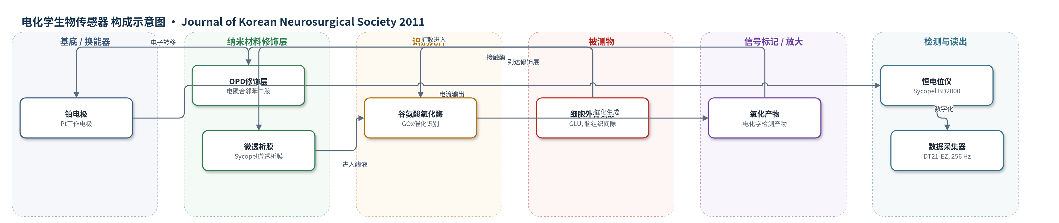
点击图片查看大图 · 依据论文自动绘制
传感器类型
电化学生物传感器
检测对象
谷氨酸(glutamate, GLU);样品基质:大鼠脑组织细胞外液(微透析液,运动皮层)
检测原理
该传感器基于微透析与酶促安培检测。大鼠运动皮层细胞外谷氨酸(GLU)经微透析膜扩散进入含谷氨酸氧化酶(GOx)的磷酸盐缓冲液(PBS)中,GOx催化谷氨酸氧化并产生可电化学检测的产物;产物在电聚合邻苯二胺(OPD)修饰的铂电极(Pt)表面发生氧化还原反应,恒电位仪记录安培电流。电流大小与透析液中谷氨酸浓度成正比,因此可实时反映脑组织细胞外谷氨酸释放水平。传感器对标准谷氨酸在50–450 µM范围内呈线性响应,灵敏度为0.22 nA/µM,R^2=0.998。
检测灵敏度
线性范围: 50-450 µM(标准谷氨酸溶液);灵敏度: 0.22 nA/µM;R^2 = 0.998
效应效果
传感器对标准谷氨酸线性响应,R^2=0.998。体内实验中,两组脑血流(CBF)和脑电图(EEG)无显著差异,说明模型一致。尼莫地平组缺血期谷氨酸最大变化67.36±2.22 µM、再灌注期75.42±4.22 µM,总释放量28520.95±2097.55,显著低于缺血组125.01±3.74 µM、133.22±2.57 µM和56994.21±1932.18;恢复时间802.00±45.75 s,短于缺血组1472.67±83.31 s。尼莫地平组海马CA1细胞存活率95.46±6.60%,缺血组47.50±5.64%(p<0.005)。作者认为缺血期输注尼莫地平可抑制谷氨酸释放并保护神经元。
传感器的构成
- 基底/换能器电极:铂电极(Pt),微透析电极内工作电极,用于安培检测。
- 修饰层:电聚合邻苯二胺(O-phenylenediamine, OPD)修饰层,0.65 V电聚合20 min,用于修饰铂电极表面并参与安培检测。
- 识别/催化元件:谷氨酸氧化酶(glutamate oxidase, GOx),催化谷氨酸氧化并产生可检测信号。
- 透析膜:微透析膜(Sycopel General 20-10-4-4),允许谷氨酸等小分子从脑组织扩散进入。
- 流动相:磷酸盐缓冲液(PBS)含GOx,0.5 µL/min灌注,维持酶活性并输送反应产物。
- 信号读出:恒电位仪(Sycopel BD2000)与DT21-EZ数据采集器,12-bit、256 Hz,记录电流信号。
中文摘要
谷氨酸是脑内关键兴奋性神经递质,其过度释放参与神经元损伤。为明确尼莫地平对谷氨酸释放的影响,本研究在大鼠十一血管闭塞(11-VO)全脑缺血模型中实时监测细胞外谷氨酸释放。12只大鼠随机分为缺血组和尼莫地平治疗组,采用微透析安培生物传感器同步检测细胞外谷氨酸水平,并同步记录脑血流(CBF)和脑电图(EEG)。缺血期间向CBF探头侧方持续输注尼莫地平(0.025 μg/100 g/min),并以Nissl染色评估神经保护效果。结果显示,缺血期谷氨酸最大浓度变化在缺血组为133.22±2.57 μM,尼莫地平组为75.42±4.22 μM(p<0.001);两组谷氨酸总释放量差异显著(p<0.001)。与假手术组相比,缺血组海马细胞存活率为47.50±5.64(p<0.005),尼莫地平组为95.46±6.60。结论:实时监测和Nissl染色结果表明,尼莫地平通过抑制细胞外谷氨酸释放,在大鼠11-VO全脑缺血模型中发挥神经元保护作用。
英文摘要
OBJECTIVE: Glutamate is a key excitatory neurotransmitter in the brain, and its excessive release plays a key role in the development of neuronal injury. In order to define the effect of nimodipine on glutamate release, we monitored extracellular glutamate release in real-time in a global ischemia rat model with eleven vessel occlusion.
METHODS: TWELVE RATS WERE RANDOMLY DIVIDED INTO TWO GROUPS: the ischemia group and the nimodipine treatment group. The changes of extracellular glutamate level were measured using microdialysis amperometric biosensor, in coincident with cerebral blood flow (CBF) and electroencephalogram. Nimodipine (0.025 µg/100 gm/min) was infused into lateral to the CBF probe, during the ischemic period. Also, we performed Nissl staining method to assess the neuroprotective effect of nimodipine.
RESULTS: During the ischemic period, the mean maximum change in glutamate concentration was 133.22±2.57 µM in the ischemia group and 75.42±4.22 µM (p<0.001) in the group treated with nimodipine. The total amount of glutamate released was significantly different (p<0.001) between groups during the ischemic period. The %cell viability in hippocampus was 47.50±5.64 (p<0.005) in ischemia group, compared with sham group. But, the %cell viability in nimodipine treatment group was 95.46±6.60 in hippocampus (p<0.005).
CONCLUSION: From the real-time monitoring and Nissl staining results, we suggest that the nimodipine treatment is responsible for the protection of the neuronal cell death through the suppression of extracellular glutamate release in the 11-VO global ischemia model of rat.