组成图示
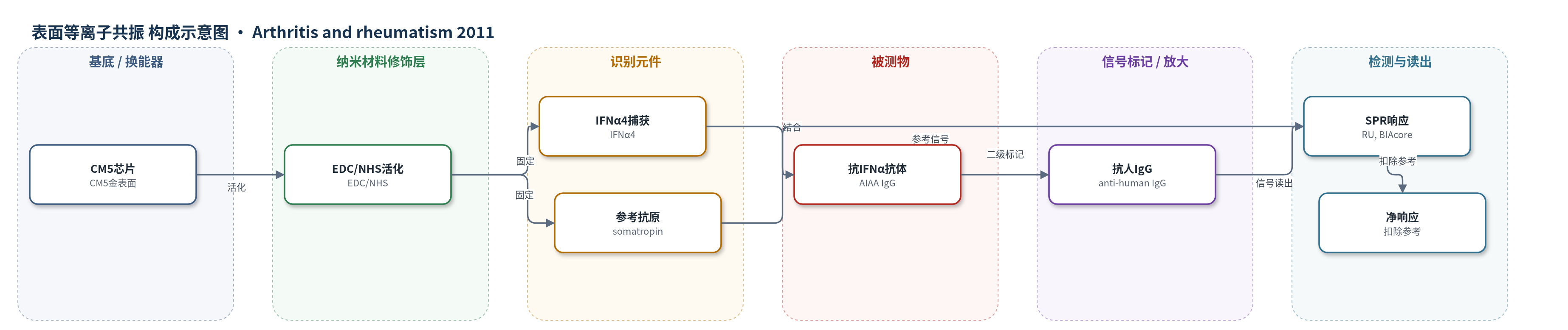
点击图片查看大图 · 依据论文自动绘制
传感器类型
表面等离子共振(SPR)生物传感器
检测对象
抗干扰素-α自身抗体(anti-IFN-α autoantibodies, AIAAs);样品基质:SLE患者血清、RA患者血清、健康供体血清。
检测原理
CM5芯片表面羧基经EDC/NHS活化后,共价固定重组人IFNα4作为捕获抗原,参考通道固定人生长激素(somatropin),并用1 M乙醇胺封闭未反应位点。稀释血清流过芯片时,血清中抗IFNα IgG自身抗体(AIAA)与固定IFNα4特异性结合,使SPR响应单位(RU)增加;随后加入抗人IgG二级抗体结合AIAA,进一步放大响应。仪器记录结合曲线,净响应为IFNα4通道减去参考通道,以扣除非特异结合。AIAA浓度或亲和力越高,净RU越大;阳性判定基于健康对照均值加1.65 SD,且抗人IgG加入后至少增加100 RU。
检测灵敏度
原文未报告LOD、线性范围、灵敏度斜率或R^2。
效应效果
SPR免疫分析在49例SLE血清中检出13例(27%)AIAA阳性,25例健康对照基本无反应,25例RA中仅1例(4%)有反应性,提示对SLE相关AIAA具有较好选择性。与ELISA相比,SPR可检出更多低亲和力AIAA,文中称ELISA仅将约10%患者判为阳性,另有17%仅SPR阳性。AIAA阳性血清在体外抑制重组IFNα2活性59%–83%,健康对照约10%;也能中和PBMC来源内源性I型干扰素。AIAA阳性患者血清I型干扰素生物活性、ISM、BAFF、抗染色质/组蛋白/核糖体P抗体更低,SLEDAI更低,C3/C4更高,提示AIAA与较低疾病活动度相关。
传感器的构成
- 基底/换能器:CM5传感器芯片(金表面羧基化)与BIAcore T100 SPR换能器,提供表面结合响应。
- 化学活化层:EDC/NHS活化CM5羧基,形成氨基反应位点用于共价固定。
- 捕获抗原层:重组人IFNα4固定于flow cell 2/3,作为AIAA捕获物;flow cell 1固定人生长激素(somatropin)作参考。
- 封闭层:1 M乙醇胺封闭未反应位点,降低非特异结合。
- 样品识别层:稀释血清中的抗IFNα IgG自身抗体(AIAA)与固定IFNα4结合。
- 信号标记层:抗人IgG抗体(The Binding Site)结合AIAA,增加SPR响应。
- 读出层:BIAcore T100记录响应单位(RU),以参考通道扣除非特异结合。
中文摘要
本研究旨在表征系统性红斑狼疮(SLE)患者内源性抗干扰素-α自身抗体(AIAA)阳性者的干扰素通路活性及血清学、临床特征。采用生物传感器免疫分析检测SLE(n=49)、类风湿关节炎(n=25)和健康对照(n=25)血清中的AIAA;用U937细胞报告基因法测定血清I型干扰素生物活性及AIAA中和活性;通过微阵列分析外周血干扰素调节基因表达,并用免疫分析检测BAFF、干扰素诱导趋化因子及其他自身抗体。结果显示,生物传感器免疫分析在27%的SLE血清中检出AIAA。无监督层次聚类将患者分为IFNlow和IFNhigh两组,两组在血清I型干扰素生物活性、干扰素调节基因表达、BAFF、抗核糖体P抗体、抗染色质抗体及AIAA状态上存在差异。多数AIAA阳性患者血清I型干扰素生物活性、下游干扰素通路活性及疾病活动度低于IFNhigh组;AIAA阳性血清在体外可有效中和I型干扰素活性。结论:SLE患者常携带AIAA,AIAA阳性者I型干扰素生物活性及下游通路活性降低,AIAA可能通过削弱IFN-α效应影响SLE临床过程。
英文摘要
OBJECTIVE: Numerous observations implicate interferon-α (IFNα) in the pathophysiology of systemic lupus erythematosus (SLE); however, the potential impact of endogenous anti-IFNα autoantibodies (AIAAs) on IFN-pathway and disease activity is unclear. The aim of this study was to characterize IFN-pathway activity and the serologic and clinical profiles of AIAA-positive patients with SLE.
METHODS: Sera obtained from patients with SLE (n = 49), patients with rheumatoid arthritis (n = 25), and healthy control subjects (n = 25) were examined for the presence of AIAAs, using a biosensor immunoassay. Serum type I IFN bioactivity and the ability of AIAA-positive sera to neutralize IFNα activity were determined using U937 cells. Levels of IFN-regulated gene expression in peripheral blood were determined by microarray, and serum levels of BAFF, IFN-inducible chemokines, and other autoantibodies were measured using immunoassays.
RESULTS: AIAAs were detected in 27% of the serum samples from patients with SLE, using a biosensor immunoassay. Unsupervised hierarchical clustering analysis identified 2 subgroups of patients, IFN(low) and IFN(high) , that differed in the levels of serum type I IFN bioactivity, IFN-regulated gene expression, BAFF, anti-ribosomal P, and anti-chromatin autoantibodies, and in AIAA status. The majority of AIAA-positive patients had significantly lower levels of serum type I IFN bioactivity, reduced downstream IFN-pathway activity, and lower disease activity compared with the IFN(high) patients. AIAA-positive sera were able to effectively neutralize type I IFN activity in vitro.
CONCLUSION: Patients with SLE commonly harbor AIAAs. AIAA-positive patients have lower levels of serum type I IFN bioactivity and evidence for reduced downstream IFN-pathway and disease activity. AIAAs may influence the clinical course in SLE by blunting the effects produced by IFNα.