组成图示
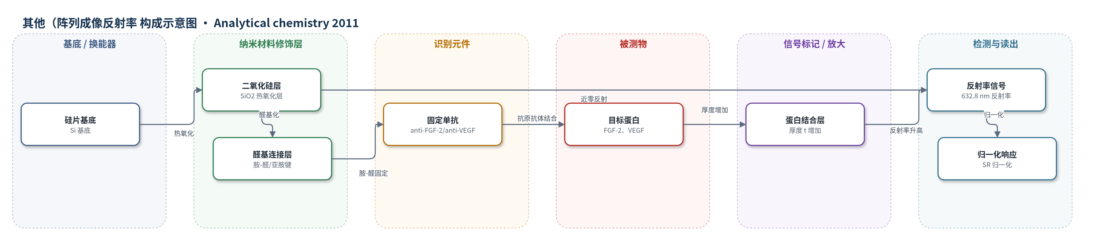
点击图片查看大图 · 依据论文自动绘制
传感器类型
其他(阵列成像反射率(AIR)生物传感器)
检测对象
成纤维细胞生长因子-2(FGF-2)、血管内皮生长因子(VEGF);样品基质:缓冲液中的蛋白溶液(文中提及可应用于血清、唾液、伤口液等,但实验为蛋白溶液/缓冲液)
检测原理
传感器利用硅片上 SiO2 与固定抗体层在 632.8 nm s 偏振、70.7° 入射下形成近零反射条件。目标蛋白与表面单抗结合后,在识别层上方形成蛋白结合层,使总光学厚度增加,破坏近零反射,反射率升高。表面覆盖度按 Langmuir 等温线随蛋白浓度变化,结合 SPR 测得的 KD 和椭偏仪测得的饱和厚度 tmax,可把浓度转换为厚度,再由特征矩阵法计算反射率。由于反射率对厚度高度非线性,低浓度下信号灵敏;归一化反射率呈 S 形,KD 对应 25% 最大响应。微阵列因抗体固定取向/表面密度不均,需双位点 Langmuir 或 Sips 等温线描述。
检测灵敏度
原文未报告 LOD、线性范围或灵敏度斜率;模型拟合 R^2:FGF-2 宏观点单点 Langmuir R^2 = 0.98;FGF-2 微阵列双位点 Langmuir/Sips R^2 = 0.98;VEGF 微阵列双位点 Langmuir/Sips R^2 = 0.99。
效应效果
该文以 SPR 和光谱椭偏仪作为互补技术交叉验证 AIR。FGF-2 宏观点数据与单点 Langmuir 模型高度一致(R^2=0.98),说明大点表面亲和力较均一;微阵列中 FGF-2 与 VEGF 的低浓度响应偏离单点模型,双位点 Langmuir 或 Sips 等温线拟合更好(FGF-2 微阵列 R^2=0.98,VEGF 微阵列 R^2=0.99),表明非定向固定导致结合位点异质性。实验为三次重复,误差棒为标准差,但未报告 RSD、选择性、稳定性、实际样品回收率或 LOD。作者认为 AIR 响应可预测,适用于不同亲和力探针-分析物体系,可推广至其他无标记生物传感平台,用于血清、唾液、伤口液中生长因子的定量检测。
传感器的构成
- 基底/换能器:平面硅片(Si),提供光学反射基底与机械支撑
- 光学修饰层:热氧化二氧化硅(SiO2)薄膜,与探针层共同形成约1420 Å近零反射光学结构
- 连接化学层:表面醛基与抗体氨基的胺-醛缩合/亚胺键(imine formation),实现非定向抗体固定
- 识别元件:单克隆抗体(anti-FGF-2、anti-VEGF),捕获目标蛋白
- 目标层:FGF-2 或 VEGF 蛋白结合层,结合后增加表面层厚度
- 信号标记物:无(label-free,不依赖荧光、酶或其他标记)
- 光学读出:632.8 nm s 偏振光、70.7°入射的成像反射率系统,测量反射率变化
中文摘要
分析技术发展的关键步骤是确认实验响应与其理论框架预测一致,从而对生物传感器进行定量验证,并将其输出信号与被测分析物浓度相关联。本文报道阵列成像反射率(AIR)这一高灵敏无标记生物传感方法的定量响应是探针和分析物性质的可预测函数。作者首先将描述表面探针-分析物相互作用的标准单点 Langmuir 结合模型纳入 AIR 厚度依赖反射率理论模型,建立分析物浓度与 AIR 响应之间的假设关系。随后,利用光谱椭偏仪、表面等离子共振(SPR)和 AIR 对两个生物医学重要蛋白——成纤维细胞生长因子-2(FGF-2)和血管内皮生长因子(VEGF)——的抗体结合进行验证。结果表明,1:1 单点 Langmuir 模型能准确描述宏观点 AIR 阵列的响应;而双点 Langmuir 模型或 Sips 等温线更能描述 AIR 微阵列的行为。研究确认了 AIR 在不同探针-分析物亲和力范围内的定量性能,所建立的方法可推广到其他无标记生物传感平台,有助于更准确地定量解释传感器响应。
英文摘要
One of the critical steps in the development of an analytical technique is to confirm that its experimental response correlates with predictions derived from the theoretical framework on which it is based. This validates the technique quantitatively and, in the case of a biosensor, facilitates a correlation of the sensor's output signal to the concentration of the analyte being tested. Herein we report studies demonstrating that the quantitative response of arrayed imaging reflectometry (AIR), a highly sensitive label-free biosensing method, is a predictable function of the probe and analyte properties. We first incorporated a standard one-site Langmuir binding model describing probe-analyte interactions at the surface into the theoretical model for thickness-dependent reflectance in AIR. This established a hypothetical correlation between the analyte concentration and the AIR response. Spectroscopic ellipsometry, surface plasmon resonance, and AIR were then used to validate this model for two biomedically important proteins, fibroblast growth factor-2 and vascular endothelial growth factor. While our studies demonstrated that the 1:1 one-site Langmuir model accurately described the observed response of macrospot AIR arrays, either a two-site Langmuir model or a Sips isotherm better described the behavior of AIR microarrays. These studies confirmed the quantitative performance of AIR across a range of probe-analyte affinities. Furthermore, the methodology developed here can be extended to other label-free biosensing platforms, thus facilitating a more accurate and quantitative interpretation of the sensor response.