组成图示
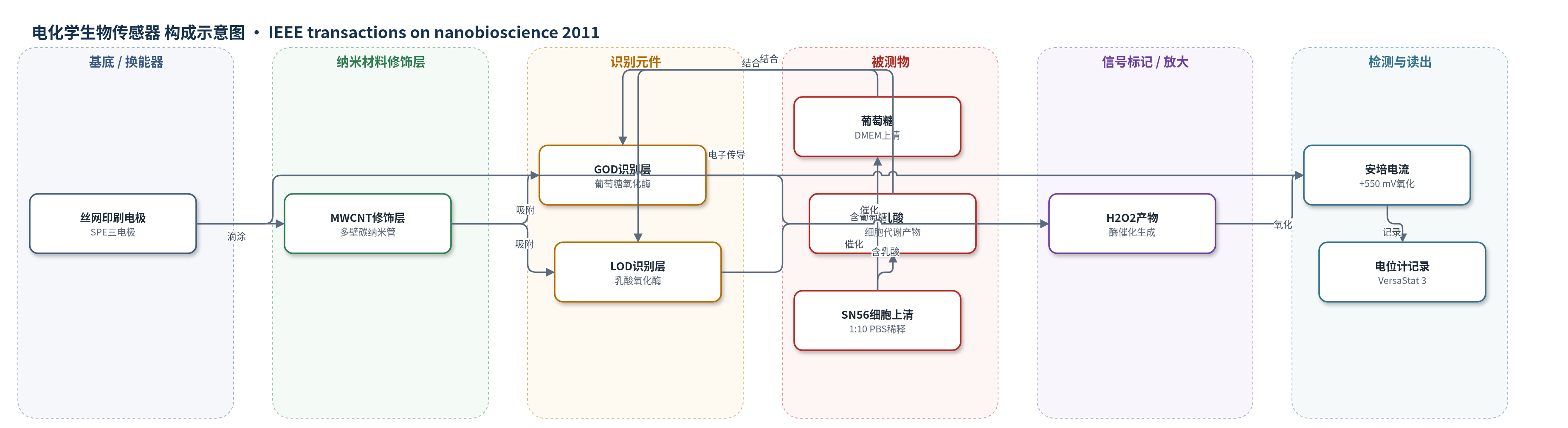
点击图片查看大图 · 依据论文自动绘制
传感器类型
电化学生物传感器
检测对象
乳酸(lactate / L-lactate)、葡萄糖(glucose / D-glucose);样品基质:SN56 细胞培养上清(DMEM,1:10 PBS 稀释),校准用 PBS/Milli-Q。
检测原理
本传感器为第二代安培型酶生物传感器。GOD或LOD吸附于MWCNT修饰的SPE工作电极表面,葡萄糖或乳酸进入酶活性位点FAD附近并被氧化,分别生成葡萄糖酸或丙酮酸,同时按1:1化学计量产生H2O2。MWCNT随机分散形成纳米结构导电网络,其侧壁和尖端场发射及高比表面积促进酶活性位点与电极之间的直接电子转移,降低界面阻抗并放大电流。在+550 mV恒电位下,H2O2在工作电极被氧化并释放电子,产生与H2O2浓度成正比的安培电流;由于酶反应化学计量关系,电流随乳酸或葡萄糖浓度增加而增加。该机制无需化学 mediator,MWCNT起到电子桥接和信号放大作用。
检测灵敏度
LOD: 28 μM(lactate)、73 μM(glucose);线性范围: 0.5-2.0 mM(lactate)、0.5-4.0 mM(glucose);灵敏度: 40.1 μA mM−1 cm−2(lactate)、27.7 μA mM−1 cm−2(glucose)
效应效果
与裸电极相比,MWCNT修饰电极将电流从nA级提高到μA级,H2O2灵敏度约提高7倍。乳酸灵敏度40.1 μA mM−1 cm−2,LOD 28 μM,线性范围0.5-2.0 mM;葡萄糖灵敏度27.7 μA mM−1 cm−2,LOD 73 μM,线性范围0.5-4.0 mM。乳酸灵敏度约为文献最高报道值的2倍,葡萄糖与文献同数量级,且未观察到交叉干扰。在SN56细胞上清(1:10 PBS稀释)中,可区分不同细胞密度4、24、48 h的乳酸产生和葡萄糖消耗;低密度乳酸近零,高密度48 h乳酸最大。作者认为可集成培养皿实现自动化细胞培养监测,适用于胚胎或间充质干细胞。
传感器的构成
- 基底/换能器电极:丝网印刷电极 SPE(DRP-110),石墨工作电极、石墨对电极、Ag/AgCl 参比电极,构成三电极体系并固定工作电位。
- 纳米材料修饰层:多壁碳纳米管 MWCNT(直径 10 nm、长 1-2 μm、90% 纯度),以氯仿溶液滴涂于工作电极,形成纳米结构导电层并促进电子转移。
- 识别元件:葡萄糖氧化酶 GOD(Aspergillus Niger,EC 1.1.3.4)或乳酸氧化酶 LOD(Pediococcus,EC 1.13.12.4),滴加吸附于 MWCNT 表面,催化底物氧化。
- 信号产物:过氧化氢 H2O2,由 GOD/LOD 催化葡萄糖或乳酸生成,在 +550 mV 工作电极氧化并释放电子,形成安培电流。
- 缓冲介质:PBS 0.01 M(pH 7.4)及 1:10 稀释 DMEM,用于酶溶解、校准和细胞培养上清检测。
中文摘要
细胞培养中代谢物监测可提供细胞系状态的实时信息,对胚胎干细胞和间充质干细胞等机制尚不明确的细胞系尤为重要,也有望推动培养皿集成化与细胞培养自动化。目前生物反应器主要监测pH、pO2、电子阻抗和温度等少数过程变量。在多种生物传感器策略中,碳纳米管具有适合高灵敏检测的特性。本文报道利用多壁碳纳米管构建纳米结构电极,用于乳酸和葡萄糖检测。通过模拟预测随机分散在电极表面的碳纳米管随取向变化的行为,并比较非纳米结构与纳米结构电极,表明若无纳米结构修饰,蛋白质与电极之间的直接电子转移无法实现。所开发生物传感器在灵敏度和检出限方面进行了表征,并与已发表结果比较。利用该传感器监测细胞培养中乳酸的产生,同时检测葡萄糖以验证乳酸行为。
英文摘要
Monitoring of metabolic compounds in cell cultures can provide real-time information of cell line status. This is particularly important in those lines not fully known, as the case of embryonic and mesenchymal cells. On the other hand, such approach can pave the way to fully automated systems for growing cell cultures, when integrated in Petri dishes. To date, the main efforts emphasize the monitoring of few process variables, like pH, pO(2), electronic impedance, and temperature in bioreactors. Among different presented strategies to develop biosensors, carbon nanotubes exhibit great properties, particularly suitable for high-sensitive detection. In this work, nanostructured electrodes by using multiwalled carbon nanotubes are presented for the detection of lactate and glucose. Some results from simulations are illustrated in order to foresee the behavior of carbon nanotubes depending on their orientation, when they are randomly dispersed onto the electrode surface. A comparison between nonnanostructured and nanostructured electrodes is considered, showing that direct electron-transfer between the protein and the electrode is not possible without nanostructuration. Such developed biosensors are characterized in terms of sensitivity and detection limit, and are compared to previously published results. Lactate production is monitored in a cell culture by using the developed biosensor, and glucose detection is also performed to validate lactate behavior.