组成图示
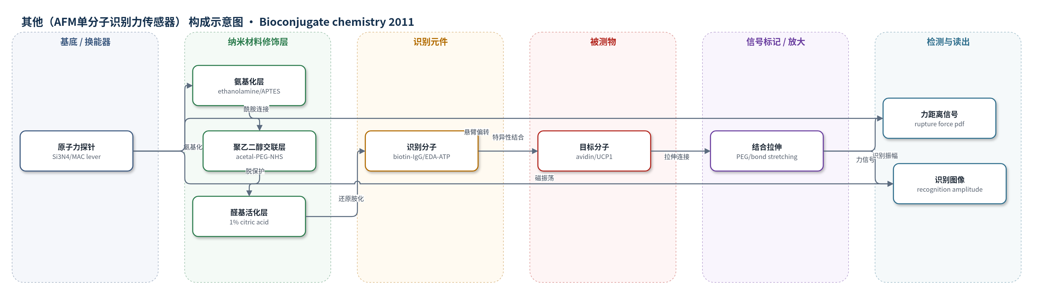
点击图片查看大图 · 依据论文自动绘制
传感器类型
其他(AFM单分子识别力传感器)
检测对象
卵白蛋白(avidin,吸附于云母表面)、解偶联蛋白1(UCP1,位于云母支撑平面磷脂双分子层中)
检测原理
AFM探针尖端经氨基化、acetal-PEG-NHS连接和温和脱保护后,形成末端苯甲醛,再与biotin-IgG或EDA-ATP的氨基经NaCNBH3还原胺化共价偶联。探针接近样品时,识别分子与靶标avidin或UCP1特异性结合;回缩时PEG链和分子间键被拉伸,达到临界力后发生断裂,使悬臂梁偏转跳变,力-距离曲线给出结合概率与断裂力。TREC模式中,磁性悬臂梁振荡,结合事件拉伸PEG并抑制向上振幅,形成识别图像;加入游离ATP竞争后识别信号消失。信号强度随靶标密度/可及性变化,无酶或核酸放大。
检测灵敏度
未报告LOD、线性范围、灵敏度斜率、相关系数
效应效果
特异性方面,游离streptavidin封闭后,avidin-biotin结合概率由21%降至1.5%(碘/丙酮法),或由32.6%降至4.4%(柠檬酸法)。芯片定量显示acetal-PEG-NHS在1 mg/mL即可高特异偶联:ethanolamine法15 min柠檬酸处理特异位点1327±185 μm−2,非特异159±49 μm−2;APTES法891±117 μm−2,非特异118±27 μm−2。封闭残余醛基未显著降低非特异结合。探针可PBS 4℃保存至3天。UCP1实验中,4.8 mM游离ATP使识别图像消失而形貌保留,证明识别来自ATP-UCP1结合。相比aldehyde-PEG-NHS需6.6 mg/mL抑制成环,新方法低浓度有效,适用于单分子识别力显微镜。
传感器的构成
- 基底/换能器:Si3N4 AFM tip或MAC magnetic-coated lever,承载探针并转换形变/力信号
- 氨基功能化层:ethanolamine hydrochloride或APTES在tip表面生成NH2,用于连接交联剂
- 柔性交联层:acetal-PEG-NHS或aldehyde-PEG-NHS,NHS端与NH2成酰胺,PEG链提供柔性
- 脱保护/活化层:1% citric acid或0.1% iodine/acetone将acetal转为benzaldehyde,暴露氨基反应位点
- 识别元件:biotin-IgG或EDA-ATP,经NaCNBH3还原胺化与benzaldehyde共价连接
- 信号标记物:无独立标记,biotin-IgG/EDA-ATP结合后直接改变AFM力/振幅信号
- 封闭剂:ethanolamine或glycine可选封闭残余benzaldehyde,降低非特异结合
- 信号读出:AFM cantilever deflection、force-distance rupture event或TREC recognition amplitude
中文摘要
原子力显微镜(AFM)探针可通过在尖端固定一个或少数生物分子升级为特异性生物传感器,用于在样品表面定位对应靶分子或研究相互作用生物物理参数。若传感器分子经柔性聚合物链连接,其功能显著增强。常规探针功能化需先在表面生成反应基团,再连接双功能交联剂,最后偶联探针分子。但探针表面最常用的氨基与许多生物分子(如抗体)可用的氨基相同,易导致交联剂与邻近氨基发生双价成环。本研究采用新交联剂acetal-PEG-NHS,其一端为活化羧基,另一端为缩醛保护的苯甲醛;活化羧基先与氨基化探针单侧快速连接,随后用1%柠檬酸处理1–10 min将缩醛温和转化为氨基反应性苯甲醛,且不损伤标准AFM探针。磁性涂层探针在1%柠檬酸中会退磁,可通过先脱保护再连接PEG交联剂解决;相应aldehyde-PEG-NHS在高浓度下可抑制双价结合。由此,磁性AFM探针可偶联ATP乙二胺衍生物,并与云母支撑平面磷脂双分子层中纯化的解偶联蛋白1(UCP1)发生特异性相互作用。
英文摘要
The measuring tip of an atomic force microscope (AFM) can be upgraded to a specific biosensor by attaching one or a few biomolecules to the apex of the tip. The biofunctionalized tip is then used to map cognate target molecules on a sample surface or to study biophysical parameters of interaction with the target molecules. The functionality of tip-bound sensor molecules is greatly enhanced if they are linked via a thin, flexible polymer chain. In a typical scheme of tip functionalization, reactive groups are first generated on the tip surface, a bifunctional cross-linker is then attached with one of its two reactive ends, and finally the probe molecule of interest is coupled to the free end of the cross-linker. Unfortunately, the most popular functional group generated on the tip surface is the amino group, while at the same time, the only useful coupling functions of many biomolecules (such as antibodies) are also NH(2) groups. In the past, various tricks or detours were applied to minimize the undesired bivalent reaction of bifunctional linkers with adjacent NH(2) groups on the tip surface. In the present study, an uncompromising solution to this problem was found with the help of a new cross-linker ("acetal-PEG-NHS") which possesses one activated carboxyl group and one acetal-protected benzaldehyde function. The activated carboxyl ensures rapid unilateral attachment to the amino-functionalized tip, and only then is the terminal acetal group converted into the amino-reactive benzaldehyde function by mild treatment (1% citric acid, 1-10 min) which does not harm the AFM tip. As an exception, AFM tips with magnetic coating become demagnetized in 1% citric acid. This problem was solved by deprotecting the acetal group before coupling the PEG linker to the AFM tip. Bivalent binding of the corresponding linker ("aldehyde-PEG-NHS") to adjacent NH(2) groups on the tip was largely suppressed by high linker concentrations. In this way, magnetic AFM tips could be functionalized with an ethylene diamine derivative of ATP which showed specific interaction with mitochondrial uncoupling protein 1 (UCP1) that had been purified and reconstituted in a mica-supported planar lipid bilayer.