组成图示
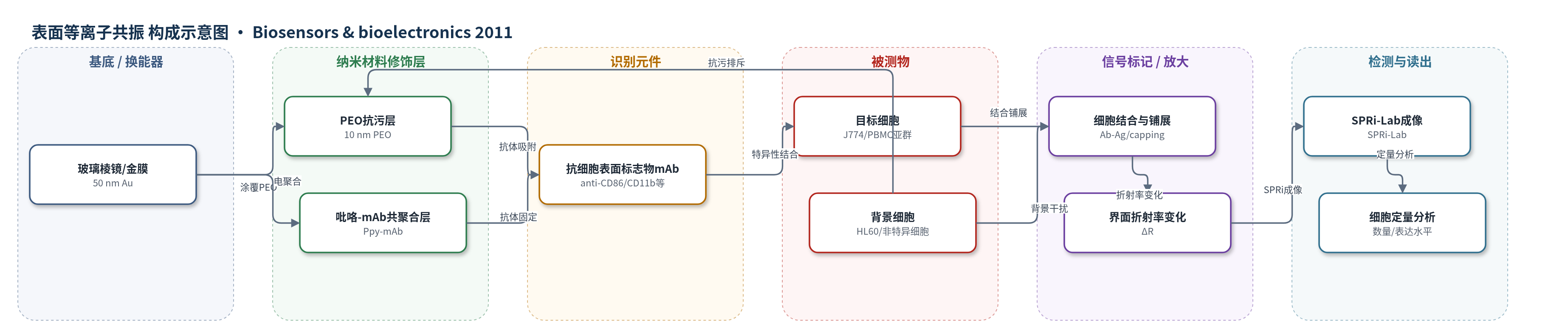
点击图片查看大图 · 依据论文自动绘制
传感器类型
表面等离子共振(SPR)生物传感器
检测对象
小鼠巨噬细胞(J774)、人早幼粒细胞(HL60)、人外周血单个核细胞(PBMC,含 CD3+ T 细胞、CD14+ 单核/巨噬细胞、CD19+ B 细胞、CD56+ NK 细胞);样品基质:PBS/BSA 细胞悬液、人外周血 PBMC 悬液
检测原理
该传感器以金膜 SPR 芯片为换能器,表面固定抗细胞表面标志物单克隆抗体(mAb)。目标细胞经抗体-抗原(Ab-Ag)相互作用被特异性捕获;受体聚集和抗原帽化使细胞在芯片表面铺展,增加约 100 nm SPR 探测深度内的界面质量与折射率变化。金膜表面等离子体共振反射率随界面折射率改变,SPRi 成像仪同步记录各抗体斑点的反射率变化 ΔR。ΔR 随结合细胞数量增加而增大,并受目标表面标志物表达密度影响。PEO 抗污层减少背景细胞非特异吸附,提高信噪比,从而降低检测阈值。
检测灵敏度
检测阈值(未涂 PEO): 约 5% 注入细胞,即 10,000 个 J774 细胞;检测阈值(PEO 涂覆): 1000 个结合细胞,相当于 0.5% 注入细胞;灵敏度提高: 10 倍。
效应效果
系统选择性良好:anti-CD86 和 anti-CD11b 斑点产生明显信号,anti-CD8、anti-CD90 及无抗体吡咯对照无信号;PEO 涂覆显著抑制非特异结合。细胞活力未受明显影响,回收细胞活力 >98%,结合细胞 24 h 后 >90% 存活。未涂 PEO 时检测阈值为约 5% 注入细胞(10,000 个 J774),PEO 涂覆后降至 1000 个结合细胞(0.5% 注入细胞),灵敏度提高 10 倍,动态范围至少一个数量级。对人 PBMC 中 3×10^4 个细胞可区分 CD3+、CD14+、CD19+、CD56+ 亚群,与流式细胞术(62%、14%、2%、1%)相关,作者认为可用于复杂临床样品稀有细胞检测。
传感器的构成
- 基底/换能器:玻璃棱镜镀 50 nm 金层(glass prism coated with 50 nm Au),提供 SPR 换能表面
- 抗污修饰层:10 nm 聚环氧乙烷薄膜(PEO,plasma-polymerized PEO),涂覆金表面,抑制蛋白与细胞非特异吸附
- 识别元件固定层:吡咯-单克隆抗体共聚合物(polypyrrole–mAb conjugate,Ppy-mAb),通过电化学共聚合将 mAb 固定于金表面;或在 PEO 表面直接吸附 mAb
- 识别元件:抗细胞表面标志物单克隆抗体(mAb,如 anti-CD86、anti-CD11b、anti-CD3、anti-CD14、anti-CD19、anti-CD56),特异性捕获目标细胞
- 封闭/抗非特异层:聚-L-赖氨酸-接枝-聚乙二醇共聚物(poly(l-lysine)-graft-(polyethylene glycol) copolymer)及 PBS/BSA 2%、Tween-20,用于封闭非特异结合
- 信号标记物:无外源标记,结合细胞本身作为质量/折射率变化源
- 信号读出:SPRi 成像仪(SPRi-Lab)与 Waters 600E 泵,监测反射率变化 ΔR
中文摘要
微阵列是细胞分离与检测的有前景工具,但尚未在生物学中广泛应用,原因在于其敏感性与复杂生物样品兼容性缺乏证明,且未证实其使用不会诱导异常细胞效应。本文表征并优化了一种将抗体微阵列与表面等离子共振成像(SPRi)相结合的技术。以小鼠巨噬细胞系 J774 为模型,证明了抗体微阵列的结合特异性,以及 SPRi 信号与结合细胞数量和细胞表面标志物表达水平之间的相关性。共聚焦显微镜显示,细胞通过抗体-抗原相互作用结合到芯片后发生形态变化,反映相关细胞表面标志物的密度,而荧光显微镜表明细胞活力未受影响。在芯片金表面沉积聚环氧乙烷(PEO)薄膜可使微阵列-SPRi 系统的检测阈值降低 10 倍,从而能够检测仅占混合群体 0.5% 的细胞。该方法还通过两个应用展示潜力:表征配体-细胞受体相互作用以确定受体特异性,以及分析人外周血单个核细胞,证明其适用于复杂生物样品分析。
英文摘要
Microarrays are promising tools for cell isolation and detection. However, they have yet to be widely applied in biology. This stems from a lack of demonstration of their sensitivity and compatibility with complex biological samples, and a lack of proof that their use does not induce aberrant cellular effects. Herein, we characterized and optimized a recently developed technology associating antibody microarrays with surface plasmon resonance imaging (SPRi). Using a murine macrophage cell line we demonstrate the binding specificity of our antibody-microarrays and the correlation between SPRi signals and both the number of bound cells, and the level of expression of cell surface markers. Confocal microscopy reveals that cell binding to the chip through antibody-antigen interactions underwent morphological changes reflecting the density of the relevant cell surface marker without affecting cell viability as shown by fluorescent microscopy. The detection threshold of the microarray-SPRi system is lowered 10-fold by applying a polyethylene oxide film to the gold surface of the chip. This increased sensitivity allows the detection of cells representing as little as 0.5% of a mixed population. The potential of this method is illustrated by two applications: characterization of ligand-cell receptor interactions, allowing determination of receptor specificity, and analysis of peripheral blood mononuclear cells, demonstrating the suitability of this tool for the analysis of complex biological samples.