组成图示
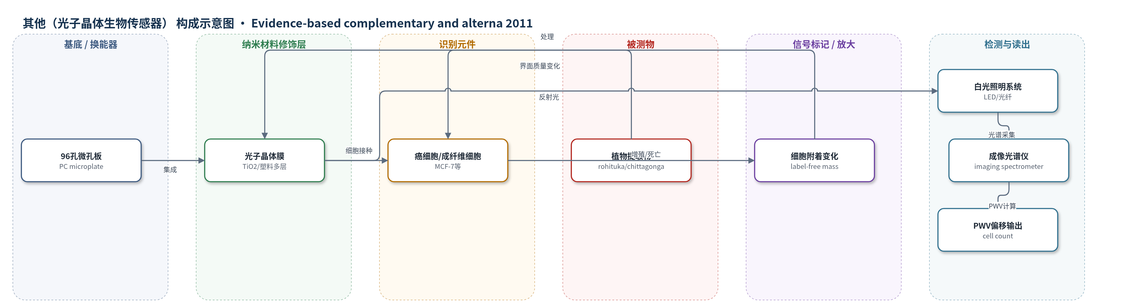
点击图片查看大图 · 依据论文自动绘制
传感器类型
其他(光子晶体生物传感器)
检测对象
Amoora rohituka 提取物、Amoora chittagonga 提取物(Pet-Ether、CH2Cl2、EtoAC、MeOH),样品基质:细胞培养液(DMEM/IMDM)中的 MCF-7、HTB-126、Panc-1、Mia-Paca2、Capan-1、Hs68 细胞培养体系。
检测原理
光子晶体由 TiO2 高折射率层与塑料低折射率层构成,白光垂直入射时产生反射峰波长值(PWV)。癌细胞或成纤维细胞接种后附着于晶体表面,表面质量与有效折射率增加,PWV 发生红移。加入不同浓度 Amoora 提取物后,若提取物诱导细胞凋亡、坏死或抑制增殖,细胞从表面脱附或数量减少,PWV 相对对照下降;若促进增殖则 PWV 增加。成像光谱仪采集各像素反射光谱,通过 PWV 变化定量细胞附着数量与存活分数,从而反映提取物细胞毒性。该方法无荧光或比色标记,信号随提取物浓度呈剂量依赖变化。
检测灵敏度
相关系数: R^2 = 0.7158、R^2 = 0.922、R^2 = 0.9094
效应效果
MTT 法显示 chittagonga 石油醚、二氯甲烷和 rohituka 石油醚对 MCF-7 的 IC50 约 42、48、41 µg/mL;chittagonga 二氯甲烷对 HTB-126、Panc-1、Mia-Paca2、Capan-1 约 43、39、30、65 µg/mL。PC 传感器对 MCF-7 的 IC50 约 40、51、38 µg/mL,与 MTT 相关系数 R^2 为 0.9094、0.7158、0.922。提取物对正常 Hs68 无毒性(IC50 > 100 µg/mL),提示癌细胞选择性。未报告 RSD、稳定性与回收率。PC 法可无标记成像、高通量筛选,作者认为适合天然化合物抗癌活性评价。
传感器的构成
- 基底/换能器:96孔标准微孔板底部集成光子晶体(photonic crystal, PC),作为光学谐振换能器。
- 光子晶体膜:高折射率 TiO2 与低折射率塑料交替层,形成反射峰波长值(peak wavelength value, PWV),对表面附着质量敏感。
- 识别元件:接种的癌细胞或成纤维细胞(MCF-7、HTB-126、Panc-1、Mia-Paca2、Capan-1、Hs68),附着于传感器表面并作为生物响应元件。
- 信号标记物:无外源标记,细胞附着/脱附本身引起光子晶体反射峰变化。
- 读出装置:LED 白光、照明光纤、检测光纤、准直透镜和成像光谱仪,测量 PWV 偏移并计算细胞计数/存活分数。
中文摘要
癌症化疗药物对正常组织毒性大,因此替代医学受到关注。Amoora rohituka 被认为具有抗肿瘤和抗菌活性。本研究将 Amoora rohituka 与 Amoora chittagonga 茎皮分别用石油醚、二氯甲烷、甲醇或乙醇(EtoAC)分级,评价其对两种乳腺癌细胞(MCF-7、HTB-126)、三种胰腺癌细胞(Panc-1、Mia-Paca2、Capan-1)及正常人包皮成纤维细胞 Hs68 的细胞毒性。采用 MTT 比色法和无标记光子晶体生物传感器成像法进行浓度系列检测。结果显示,MCF-7 细胞中 chittagonga 石油醚和二氯甲烷提取物以及 rohituka 石油醚提取物诱导细胞毒性,而 chittagonga EtoAC 和 rohituka 甲醇提取物无毒性;HTB-126、Panc-1、Mia-Paca2 和 Capan-1 中仅 chittagonga 二氯甲烷提取物显示显著细胞毒性。所有提取物对 Hs68 正常成纤维细胞无毒性,提示其可能具有癌细胞选择性。结果支持进一步研究 Amoora 物种的抗癌活性并鉴定活性成分。
英文摘要
Chemotherapeutic agents for cancer are highly toxic to healthy tissues and hence alternative medicine avenues are widely researched. Majority of the recent studies on alternative medicine suggested that Amoora rohituka possesses considerable antitumor and antibacterial properties. In this work, rohituka and chittagonga, fractionated with petroleum ether, dichloromethane, and ethanol, were explored for their anticancer potential against two breast cancer (MCF-7 and HTB-126) and three pancreatic cancer (Panc-1, Mia-Paca2, and Capan1). The human foreskin fibroblast, Hs68, was also included. Cytotoxicity of each extract was analyzed using the MTT assay and label-free photonic crystal biosensor assay. A concentration series of each extract was performed on the six cell lines. For MCF-7 cancer cells, the chittagonga (Pet-Ether and CH(2)Cl(2)) and rohituka (Pet-Ether) extracts induced cytotoxicity; the chittagonga (EtoAC) and rohituka (MeOH) extracts did not induce cytotoxicity. For HTB126, Panc-1, Mia-Paca2, and Capan-1 cancer cells, only the chittagonga CH(2)Cl(2) extract showed a significant cytotoxic effect. The extracts were not cytotoxic to normal fibroblast Hs68 cells, which may be correlated to the specificity of Amoora extracts in targeting cancerous cells. Based on these results, further examination of the potential anticancer properties Amoora species and the identification of the active ingredients of these extracts is warranted.