组成图示
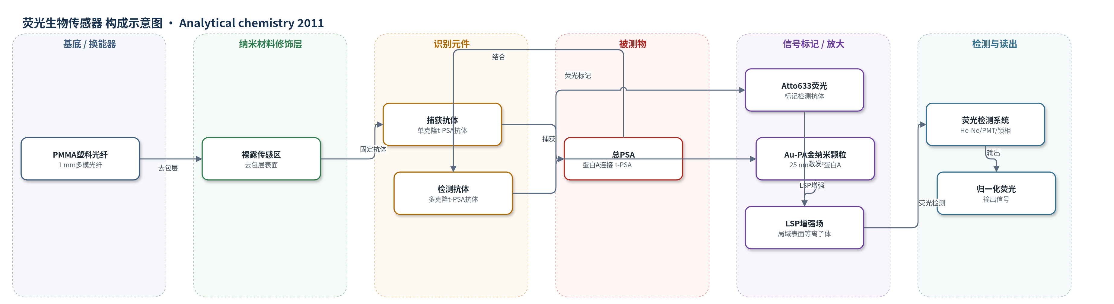
点击图片查看大图 · 依据论文自动绘制
传感器类型
荧光生物传感器
检测对象
总前列腺特异性抗原(total PSA, t-PSA);样品基质:PBS、10倍稀释健康女性血清、临床女性血清(乳腺癌组与非癌组)
检测原理
PMMA光纤裸露表面通过化学吸附固定单克隆t-PSA捕获抗体,并用BSA封闭非特异位点。加入含t-PSA的样品后,t-PSA被捕获抗体特异性结合。随后加入LSPCF探针,即Atto633标记的多克隆t-PSA检测抗体通过蛋白A连接25 nm金纳米颗粒,形成捕获抗体–t-PSA–检测抗体夹心复合物。632.8 nm He-Ne激光激发光纤传感区,金纳米颗粒产生局域表面等离子体(LSP),增强Atto633附近的局部电磁场,使荧光发射增强。侧向荧光经640 nm长通滤光片由PMT检测,并用锁相放大和归一化算法降低噪声。t-PSA浓度越高,形成的夹心复合物越多,归一化荧光信号越强,从而实现定量检测。
检测灵敏度
LOD: 0.4 pg/mL(PBS);1.8 pg/mL(10倍稀释健康女性血清);约18 pg/mL(实际血清);线性范围: 0.1–1000 pg/mL(摘要);1–1000 pg/mL(PBS);0–1000 pg/mL(10倍稀释健康女性血清);分析灵敏度: 1.4 pg/mL(PBS);2.3 pg/mL(10倍稀释健康女性血清);约23 pg/mL(实际血清);R^2 = 0.9574(PBS);R^2 = 0.9142(10倍稀释健康女性血清);拟合方程: y = 18.84 log x + 33.26(PBS);y = 20.30 log x + 14.25(10倍稀释健康女性血清)
效应效果
在PBS和10倍稀释健康女性血清中,t-PSA与归一化荧光信号线性,R^2分别为0.9574和0.9142。8例乳腺癌与8例非癌血清中,乳腺癌组均值/中位数为155.2/145.7 AU,非癌组为46.6/37.1 AU。经验AUC为0.9063,拟合AUC为0.9166;拟合灵敏度82.43%、特异性87.67%。阈值107 AU时灵敏度75%、特异性100%;93.91 AU时87.5%和75%;34.81 AU时100%和50%。作者认为其灵敏度/特异性优于乳腺球蛋白、bFGF、抗恶性抗体,并与PSA前列腺癌诊断(73.2%/85.4%)及临床检查、钼靶(54%/94%、75%/92%)相当或更优,可用于乳腺癌早期判别。
传感器的构成
- 基底/换能器:PMMA多模塑料光纤(直径1 mm),去除包层后裸露区作为传感界面,传导激发光并侧向收集荧光。
- 识别元件:单克隆t-PSA捕获抗体(5 μg/mL),化学吸附固定于光纤裸露表面,特异性捕获t-PSA。
- 封闭层:BSA(10 mg/mL,PBS),封闭未结合位点,降低非特异吸附。
- 检测识别元件:多克隆t-PSA检测抗体,与t-PSA结合形成夹心免疫复合物。
- 荧光标记物:Atto633(Lightning-Link Atto633),标记检测抗体,提供荧光信号。
- 纳米增强层:Au-PA(25 nm金纳米颗粒偶联蛋白A),结合荧光标记检测抗体,通过LSP增强局部电磁场。
- 信号读出:He-Ne激光(632.8 nm)、长通滤光片(640 nm)、PMT与锁相放大器,检测归一化荧光信号。
中文摘要
前列腺特异性抗原(PSA)被认为是乳腺癌的潜在生物标志物。正常女性血清中PSA浓度约为1 pg/mL,通常难以用现有方法检测,因此需要超灵敏检测女性血清中的PSA以区分正常乳腺疾病与恶性乳腺疾病。为提高常规免疫分析检测血清PSA的灵敏度,作者采用局域表面等离子体耦合荧光光纤生物传感器(LSPCF-FOB),将夹心免疫分析与局域表面等离子体技术相结合。在磷酸盐缓冲液(PBS)中,总PSA(t-PSA)浓度0.1–1000 pg/mL与归一化荧光信号呈线性关系,相关系数为0.9574;在10倍稀释健康女性血清中,加入的t-PSA在类似范围内也呈线性,相关系数为0.9142。临床血清检测显示,乳腺癌组归一化荧光信号均值和中位数分别为155.2和145.7,均高于非癌组的46.6和37.1。t-PSA检测的受试者工作特征曲线下面积(AUC)为0.9063,表明该方法具有较好的乳腺癌判别能力。
英文摘要
Prostate-specific antigen (PSA) has been reported to be a potential biomarker of breast cancer. Serum PSA of normal women is around 1 pg/mL, which is usually undetectable by current assay methods; thus an ultrasensitive measurement of PSA expression in women's serum is necessary to distinguish normal from malignant breast diseases. To enhance the sensitivity of conventional immunoassay technology for the detection of PSA in sera, we adopted a localized surface plasmon coupled fluorescence fiber-optic biosensor, which combines a sandwich immunoassay with the localized surface plasmon technique. The concentration of total PSA (t-PSA) (from 0.1 to 1000 pg/mL) in phosphate-buffered saline solution and the normalized fluorescence signal exhibit a linear relationship where the correlation coefficient is 0.9574. In addition, the concentration of additional t-PSA in 10-fold-diluted healthly women's serum across a similar range was measured. The correlation coefficient for this measurement is 0.9142. In clinical serum samples, moreover, the experimental results of t-PSA detection show that both the mean value and median of normalized fluorescence signals in the breast cancer group (155.2 and 145.7, respectively) are higher than those in the noncancer group (46.6 and 37.1, respectively). We also examined the receiver operating characteristic curve for t-PSA, and the area under the curve (AUC) is estimated to be 0.9063, the AUC being used to measure the performance of a test to correctly identify diseased and nondiseased subjects.