组成图示
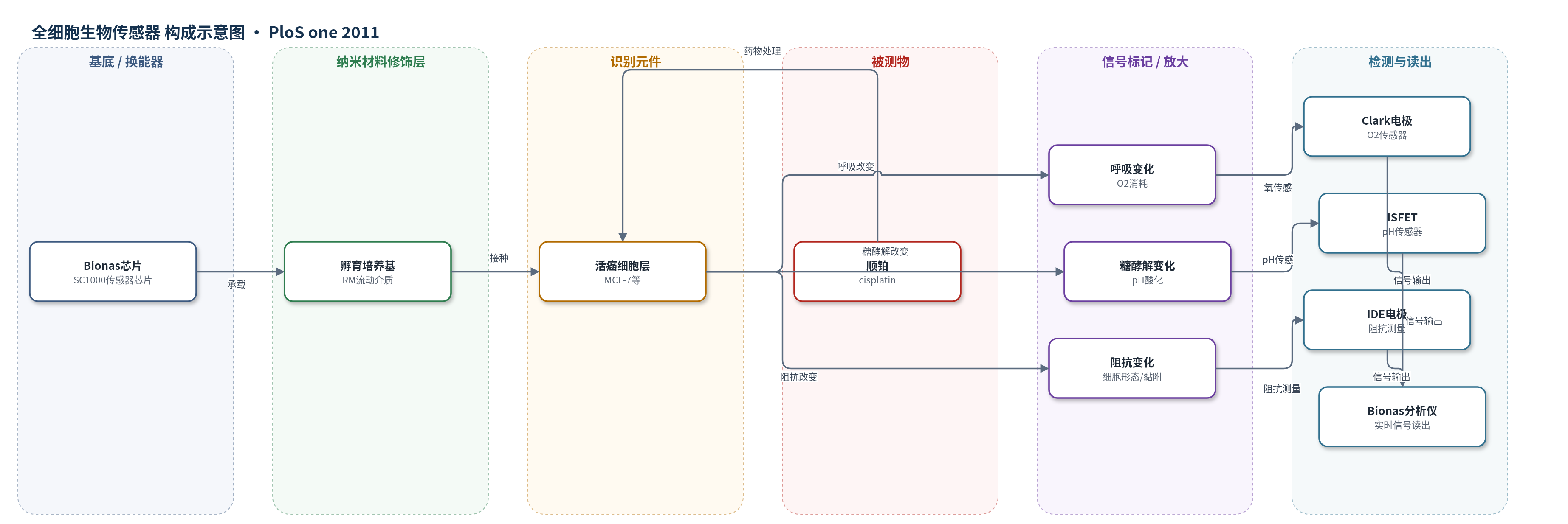
点击图片查看大图 · 依据论文自动绘制
传感器类型
全细胞生物传感器
检测对象
顺铂诱导的细胞死亡与代谢响应(cisplatin-induced cell death / cellular metabolic response);样品基质:活癌细胞培养液(DMEM培养基中的MCF-7、HT-29、HCT-116、HepG2、MDA-MB-231细胞)
检测原理
顺铂进入细胞后与DNA形成加合物,引发DNA损伤、线粒体功能障碍和应激/凋亡信号,使贴附于芯片表面的活细胞发生代谢与形态改变。细胞呼吸变化改变培养基氧浓度,由Clark型电极检测;糖酵解产生的酸化改变细胞外pH,由ISFET检测;细胞收缩、黏附丧失和膜完整性破坏改变细胞层电容/阻抗,由互指电极检测。信号随顺铂浓度和时间变化:浓度越高,呼吸下降、糖酵解改变和阻抗下降越快越明显;阻抗骤降标志细胞死亡开始。该体系无外加酶或核酸放大,依靠全细胞响应实现时间分辨读出。
检测灵敏度
未报告LOD、线性范围、灵敏度斜率或R^2。
效应效果
芯片可连续非侵入监测癌细胞对顺铂的实时响应。50 μM下,MCF-7呼吸5–6 h下降,糖酵解8–9 h升高,阻抗10–11 h下降;HT-29、HCT-116、HepG2呼吸立即下降,糖酵解6–10 h下降,阻抗8–11 h下降;MDA-MB-231仅高浓度呼吸下降,糖酵解和阻抗不变。24 h撤药后敏感细胞无恢复。分离线粒体显示顺铂4–5 h内不直接抑制呼吸,ROS无显著升高。基因芯片显示1338个探针集变化(335上调、1003下调),p53、细胞周期和凋亡富集(p=0.0023、0.001、0.0058),支持约10 h转向凋亡。
传感器的构成
- 芯片基底/换能器:Bionas 2500传感器芯片SC1000(原文未给出具体基底材料),集成孵育腔与微电极,提供连续监测平台
- 氧换能层:Clark-type电极,检测培养基中氧消耗,反映细胞呼吸
- pH换能层:ISFET(ion-sensitive field effect transistor),检测细胞外酸化,反映糖酵解
- 阻抗换能层:IDESs(interdigitated electrode structures),测量细胞层阻抗,反映细胞形态、黏附和膜功能
- 识别/响应元件:活癌细胞(MCF-7、HT-29、HCT-116、HepG2、MDA-MB-231),接种于芯片表面,作为全细胞识别元件
- 流动介质:运行培养基RM(DMEM without carbonate buffer、1 mM HEPES、0.1% FCS、1 g/L glucose),维持细胞并输送顺铂
- 刺激/分析物:cisplatin(顺铂),以不同浓度加入培养基,诱导细胞代谢与死亡响应
中文摘要
顺铂自20世纪70年代临床应用以来,其抗肿瘤机制研究广泛,但顺铂如何随时间诱导细胞死亡仍不清楚。本研究采用新型细胞生物传感器芯片系统,对多种癌细胞系进行顺铂处理,并实时连续监测细胞呼吸、糖酵解和阻抗。结果显示,所有顺铂处理细胞系中呼吸首先受到影响,随后糖酵解在HT-29、HCT-116、HepG2和MCF-7细胞中发生显著延迟性干扰,而顺铂耐药细胞系MDA-MB-231未出现糖酵解干扰。最显著的是,所有顺铂敏感细胞系在治疗后8–11 h内开始死亡,表明从药物暴露、初始响应到细胞命运决定存在明确时间框架。作者选择MCF-7细胞最显著变化时间点,直接从传感器芯片取样分析信号转导磷酸化和基因表达。该在线细胞生物传感器首次揭示顺铂处理下从代谢响应到细胞死亡开始的时间尺度,并与p53介导的细胞命运决定模型一致。
英文摘要
Since the discovery of cisplatin more than 40 years ago and its clinical introduction in the 1970s an enormous amount of research has gone into elucidating the mechanism of action of cisplatin on tumor cells. With a novel cell biosensor chip system allowing continuous monitoring of respiration, glycolysis, and impedance we followed cisplatin treatment of different cancer cell lines in real-time. Our measurements reveal a first effect on respiration, in all cisplatin treated cell lines, followed with a significant delay by interference with glycolysis in HT-29, HCT-116, HepG2, and MCF-7 cells but not in the cisplatin-resistant cell line MDA-MB-231. Most strikingly, cell death started in all cisplatin-sensitive cell lines within 8 to 11 h of treatment, indicating a clear time frame from exposure, first response to cisplatin lesions, to cell fate decision. The time points of most significant changes were selected for more detailed analysis of cisplatin response in the breast cancer cell line MCF-7. Phosphorylation of selected signal transduction mediators connected with cellular proliferation, as well as changes in gene expression, were analyzed in samples obtained directly from sensor chips at the time points when changes in glycolysis and impedance occurred. Our online cell biosensor measurements reveal for the first time the time scale of metabolic response until onset of cell death under cisplatin treatment, which is in good agreement with models of p53-mediated cell fate decision.