组成图示
点击图片查看大图 · 依据论文自动绘制
传感器类型
其他(超极化129Xe NMR生物传感器)
检测对象
转铁蛋白受体(transferrin receptor, TfR)及转铁蛋白-受体结合/内吞事件;样品基质:K562细胞悬液(PBS)
检测原理
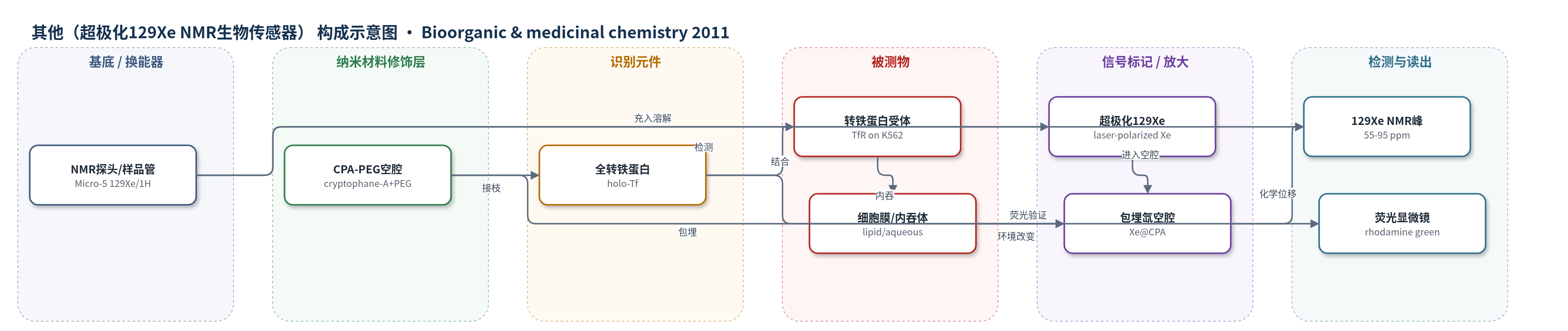
该传感器以全转铁蛋白(holo-Tf)为识别元件,与K562细胞表面转铁蛋白受体(TfR)结合并触发内吞。激光极化129Xe气体溶解于PBS/细胞悬液后,氙原子进入接枝在蛋白上的隐环烷-A(CPA)空腔;空腔微环境使包埋氙产生特征化学位移,水相约68–69 ppm,脂质/膜相约79 ppm。TfR介导的结合与内吞使传感器在细胞膜和内吞体中富集,改变Xe@CPA峰强度及水相/膜相峰比例;pronase灭活TfR后信号显著下降,证明受体依赖性。CPA疏水性也会引起非特异膜结合,BSA对照用于区分。最终通过129Xe NMR选择性激发55–95 ppm峰积分读出,荧光显微镜辅助验证。
检测灵敏度
LOD: 80 nM(约 50 pmol,使用 1 mL 气态超极化氙,直接观测法)
效应效果
K562细胞表面约1.6×10^5个TfR/细胞。5 μM B1与1.4×10^8个细胞孵育1 h后,129Xe NMR出现水相68–69 ppm和膜相79 ppm峰,峰面积总和守恒,流程稳健。pronase灭活TfR后膜相峰几乎消失,荧光无摄取,证明受体依赖。BSA对照显示CPA疏水导致非特异膜结合,但B1比B3多33%细胞相关信号,水相/膜相峰比0.59对0.28,提示特异性内吞。holo-Tf 90 μM弛豫时间147±10 s,5 μM下不影响检测。作者认为首次实现活细胞内超极化氙生物事件探测。
传感器的构成
- 识别元件:全转铁蛋白(holo-transferrin, holo-Tf),与细胞表面转铁蛋白受体(TfR)结合并介导内吞
- 氙结合空腔:隐环烷-A(cryptophane-A, CPA)衍生物,包埋129Xe并产生特征NMR化学位移
- 连接臂:聚乙二醇(PEG)间隔臂,连接CPA与蛋白,提供柔性并减少横向弛豫展宽
- 接枝活化基团:五氟苯酚酯(pentafluorophenol ester),与蛋白赖氨酸/N端氨基反应实现非特异性共价接枝
- 荧光标记物:罗丹明绿(rhodamine green),用于双模态传感器B2/B4的荧光显微镜验证
- 对照蛋白:牛血清白蛋白(BSA),用于非特异性膜结合/胞饮对照
- 超极化氙源:激光极化129Xe气体,提供高极化NMR信号
- 读出装置:Bruker Avance II 500 MHz NMR探头(Micro-5),检测包埋氙信号
中文摘要
为了在体外检测生物事件,基于超极化129Xe核磁共振的生物传感器若能弥补灵敏度差距,将成为有力工具。本文提出将隐环烷(cryptophane)前体非选择性地接枝到全转铁蛋白上构建传感器。选择该系统是因为细胞表面存在大量受体,且内吞作用可进一步提高局部密度。通过荧光显微镜和129Xe NMR研究这些生物传感器与K562细胞悬液的相互作用,显示其具有强相互作用,并表现出有趣特性:即使生物传感器被内吞,氙仍能进入隐环烷空腔并保持较高极化水平。尽管由于隐环烷疏水特性将传感器吸引至细胞膜,导致对转铁蛋白受体缺乏特异性,但这些传感器实现了首次利用超极化氙在细胞内探测生物事件。
英文摘要
For detection of biological events in vitro, sensors using hyperpolarized (129)Xe NMR can become a powerful tool, provided the approach can bridge the gap in sensitivity. Here we propose constructs based on the non-selective grafting of cryptophane precursors on holo-transferrin. This biological system was chosen because there are many receptors on the cell surface, and endocytosis further increases this density. The study of these biosensors with K562 cell suspensions via fluorescence microscopy and (129)Xe NMR indicates a strong interaction, as well as interesting features such as the capacity of xenon to enter the cryptophane even when the biosensor is endocytosed, while keeping a high level of polarization. Despite a lack of specificity for transferrin receptors, undoubtedly due to the hydrophobic character of the cryptophane moiety that attracts the biosensor into the cell membrane, these biosensors allow the first in-cell probing of biological events using hyperpolarized xenon.