组成图示
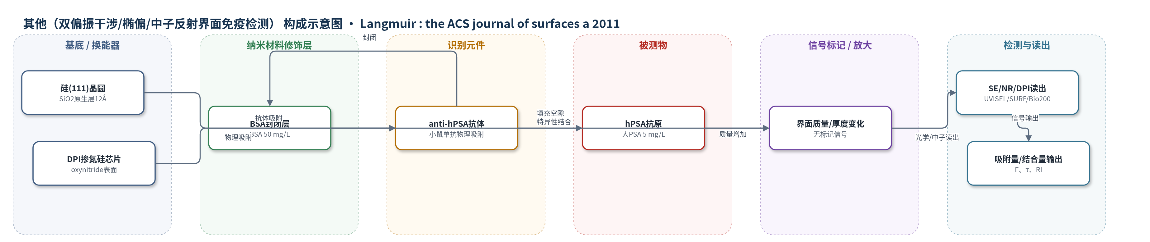
点击图片查看大图 · 依据论文自动绘制
传感器类型
其他(双偏振干涉/椭偏/中子反射界面免疫检测)
检测对象
人前列腺特异性抗原(human prostate-specific antigen, hPSA);样品基质:磷酸盐缓冲液(20 mM, pH 7)
检测原理
anti-hPSA单抗从磷酸盐缓冲液物理吸附于SiO2/水界面,低覆盖时呈平躺取向,形成约4 nm抗体层;BSA封闭空隙以抑制非特异吸附。hPSA与抗体Fab位点特异性结合后插入抗体层内部,使界面质量、散射长度密度和DPI测得的层质量/密度增加,而层厚基本保持42±2 Å。SE通过椭偏参数变化反映吸附质量,NR通过反射曲线拟合获得厚度、体积分数和表面吸附量,DPI通过双偏振干涉实时获得厚度、质量、密度和折射率。信号随hPSA结合量增加而增强,但抗体表面堆积增加会造成空间位阻,使结合效率下降,因此存在最佳抗体覆盖量。
检测灵敏度
未报告LOD、线性范围、灵敏度斜率或相关系数。
效应效果
该体系为无标记界面免疫检测,未报告LOD、线性范围、RSD或实际血清回收率。选择性来自anti-hPSA单抗对hPSA的特异性识别,BSA封闭可抑制非特异吸附;缓冲液洗涤后抗体脱附可忽略。稳定性方面,固定于SiO2表面的抗体在室温干燥保存4个月后仍保持活性,捕获抗原量为0.18±0.02 mg/m2,结合比率为0.36±0.02。NR和DPI显示抗原结合后层厚保持42±2 Å,说明抗原插入抗体层。作者认为该结果有助于优化抗体界面固定、提高生物传感器活性和稳定性,适用于未来PSA无标记传感。
传感器的构成
- 基底/换能器(SE/NR):硅(111)晶圆及原生二氧化硅(SiO2)层,厚度12±2 Å,提供抗体物理吸附界面。
- DPI换能芯片:掺氮硅/氮化硅表面,用于双偏振干涉(DPI)实时监测吸附层厚度、质量、密度和折射率。
- 识别元件:小鼠单克隆抗人PSA抗体(anti-hPSA),从磷酸盐缓冲液物理吸附于SiO2/水界面,特异性结合hPSA。
- 封闭剂:牛血清白蛋白(BSA),50 mg/L,填充抗体层空隙并阻断非特异性吸附。
- 被测物:人前列腺特异性抗原(hPSA),5 mg/L,与界面固定抗体结合。
- 信号标记物:无标记(label-free),不添加荧光/酶/纳米标记,依靠界面质量/厚度变化产生信号。
- 读出装置:光谱椭偏(SE)、中子反射(NR)和双偏振干涉(DPI),分别监测界面吸附量、层厚/散射长度密度和实时厚度/质量/折射率。
中文摘要
抗体取向及其界面抗原结合效率对免疫分析和生物传感器应用尤为重要。本文采用光谱椭偏(SE)、中子反射(NR)和双偏振干涉(DPI)研究小鼠单克隆抗人前列腺特异性抗原(anti-hPSA)抗体在二氧化硅/水界面的界面组装及后续抗原结合。结果表明,界面吸附抗体的质量密度随溶液浓度和吸附时间增加,而抗原结合效率在所研究浓度范围内随界面抗体量增加持续下降;当表面吸附抗体量约为1.5 mg/m2时,结合抗原量达到最大。该现象可用界面结构堆积或拥挤解释。NR显示,Y形抗体在低表面质量密度下平躺于界面,厚度约40 Å,相当于抗体短轴长度;此范围内松散堆积有利于抗原结合,而表面吸附量进一步增加会导致抗体片段拥挤或重叠,因空间位阻降低抗原结合。NR和DPI原位研究表明,抗原插入抗体层内部,而非在顶部形成额外层。稳定性测试显示,固定于二氧化硅表面的抗体在4个月监测期内保持稳定和活性。这些结果有助于理解抗体界面行为,并可用于生物传感器开发中活性和稳定性的控制。
英文摘要
Antibody orientation and its antigen binding efficiency at interface are of particular interest in many immunoassays and biosensor applications. In this paper, spectroscopic ellipsometry (SE), neutron reflection (NR), and dual polarization interferometry (DPI) have been used to investigate interfacial assembly of the antibody [mouse monoclonal anti-human prostate-specific antigen (anti-hPSA)] at the silicon oxide/water interface and subsequent antigen binding. It was found that the mass density of antibody adsorbed at the interface increased with solution concentration and adsorption time while the antigen binding efficiency showed a steady decline with increasing antibody amount at the interface over the concentration range studied. The amount of antigen bound to the interfacial immobilized antibody reached a maximum when the surface-adsorbed amount of antibody was around 1.5 mg/m(2). This phenomenon is well interpreted by the interfacial structural packing or crowding. NR revealed that the Y-shaped antibody laid flat on the interface at low surface mass density with a thickness around 40 Å, equivalent to the short axial length of the antibody molecule. The loose packing of the antibody within this range resulted in better antigen binding efficiency, while the subsequent increase of surface-adsorbed amount led to the crowding or overlapping of antibody fragments, hence reducing the antigen binding due to the steric hindrance. In situ studies of antigen binding by both NR and DPI demonstrated that the antigen inserted into the antibody layer rather than forming an additional layer on the top. Stability assaying revealed that the antibody immobilized at the silica surface remained stable and active over the monitoring period of 4 months. These results are useful in forming a general understanding of antibody interfacial behavior and particularly relevant to the control of their activity and stability in biosensor development.