组成图示
点击图片查看大图 · 依据论文自动绘制
传感器类型
综述或非传感器论文
检测对象
F-actin(filamentous actin,丝状肌动蛋白);样品基质:活体斑马鱼胚胎(zebrafish embryo)Rohon-Beard感觉神经元
检测原理
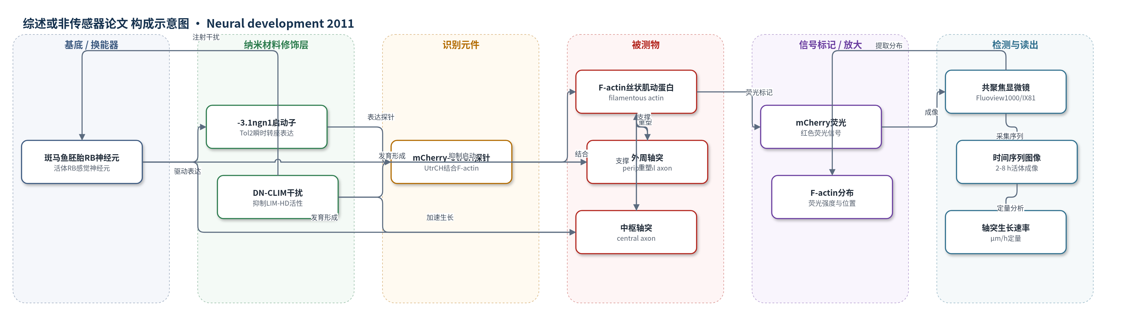
mCherry-UtrCH由utrophin的calponin同源结构域(UtrCH)与mCherry融合组成。UtrCH选择性结合F-actin,但不稳定化或显著改变其动态,因此F-actin富集、聚合或突出部位会呈现mCherry荧光增强和空间聚集。通过-3.1ngn1启动子在斑马鱼RB神经元中瞬时表达该探针,共聚焦显微镜采集Z-stack时间序列,将F-actin分布转换为荧光强度、位置和持续时间等可读信号。当外周轴突起始或分支发生时,局部F-actin先形成斑块/丝状伪足,随后探针荧光在伪足和生长锥富集;DN-CLIM抑制LIM-HD后,比较F-actin突出数量、轴突生长速率和分支数,从而判断转录因子是否影响F-actin动态或轴突定位。
检测灵敏度
未报告
效应效果
mCherry-UtrCH探针未明显干扰RB神经元运动:荧光对照与探针组中枢轴突生长速率23.8±1.4 vs 22.4±1.8 μm/h(P=0.55),外周轴突40.9±2.7 vs 40.6±2.5 μm/h(P=0.95),丝状伪足40.9±2.4 vs 37.1±2.3个/h(P=0.28)。DN-CLIM胚胎中68%神经元外周轴突未起始,20%形成短暂突起后回缩,12%成功延伸但生长更慢、分支更少;中枢轴突phase II生长速率显著加快。F-actin突出活动无显著差异(37.1±2.3 vs 33.8±1.9个/h,P=0.28),但部分神经元出现异位F-actin聚集。外周轴突启动位点平均距细胞体中心10.7±0.67 μm,67%在体节边界出脊髓。方法支持2-8 h、1-2 min间隔活体成像,揭示LIM-HD差异调控轴突行为。
传感器的构成
- 活体基底:斑马鱼胚胎(zebrafish embryo)Rohon-Beard(RB)感觉神经元,提供F-actin分布的体内环境
- 表达调控层:-3.1 ngn1顺式调控元件(-3.1ngn1)与Tol2转座系统,驱动探针在RB神经元中瞬时镶嵌表达
- 识别元件:utrophin的calponin homology结构域(UtrCH),选择性结合F-actin(filamentous actin)
- 信号标记物:mCherry荧光蛋白,与UtrCH融合为mCherry-UtrCH,在F-actin富集处产生红色荧光
- 对照标记:GFP-CAAX或TagRFP-CAAX膜靶向荧光蛋白,用于标记RB神经元膜和轴突形态
- 干扰元件:DN-CLIM(dominant negative cofactor of LIM)mRNA,抑制LIM-HD转录因子活性以比较轴突行为
- 读出系统:Olympus Fluoview1000/IX81共聚焦显微镜,采集Z-stack时间序列荧光图像
中文摘要
神经元特定形态的建立需要精确调控轴突形成、延伸和分支等细胞运动过程,而丝状肌动蛋白(F-actin)细胞骨架的动态重塑是这些过程的核心,但其在完整胚胎体内的调控机制仍不清楚。斑马鱼Rohon-Beard(RB)感觉神经元具有中枢轴突和外周轴突两种不同轨迹,LIM同源域(LIM-HD)转录因子活性对外周RB轴突形成必不可少。本研究利用活体成像观察RB神经元细胞行为及F-actin分布,采用含utrophin calponin同源结构域的F-actin生物传感器mCherry-UtrCH,在RB神经元中瞬时镶嵌表达,实时显示轴突起始、巩固和分支过程中的F-actin动态。结果显示,外周轴突从特定细胞区室起始,F-actin聚集和突出活动先于外周轴突形成;DN-CLIM抑制LIM-HD转录活性后,外周轴突起始、生长和分支受到抑制,而中枢轴突生长速率增加。进一步成像表明,LIM-HD活性并非F-actin突出活动或F-actin聚集所必需,但可影响F-actin聚集位置和轴突形成位置。该研究首次在完整胚胎中成像F-actin动态,揭示了转录因子差异调控同一神经元两条轴突形成与生长的细胞机制。
英文摘要
BACKGROUND: Development of specific neuronal morphology requires precise control over cell motility processes, including axon formation, outgrowth and branching. Dynamic remodeling of the filamentous actin (F-actin) cytoskeleton is critical for these processes; however, little is known about the mechanisms controlling motile axon behaviors and F-actin dynamics in vivo. Neuronal structure is specified in part by intrinsic transcription factor activity, yet the molecular and cellular steps between transcription and axon behavior are not well understood. Zebrafish Rohon-Beard (RB) sensory neurons have a unique morphology, with central axons that extend in the spinal cord and a peripheral axon that innervates the skin. LIM homeodomain (LIM-HD) transcription factor activity is required for formation of peripheral RB axons. To understand how neuronal morphogenesis is controlled in vivo and how LIM-HD transcription factor activity differentially regulates peripheral versus central axons, we used live imaging of axon behavior and F-actin distribution in vivo.
RESULTS: We used an F-actin biosensor containing the actin-binding domain of utrophin to characterize actin rearrangements during specific developmental processes in vivo, including axon initiation, consolidation and branching. We found that peripheral axons initiate from a specific cellular compartment and that F-actin accumulation and protrusive activity precede peripheral axon initiation. Moreover, disruption of LIM-HD transcriptional activity has different effects on the motility of peripheral versus central axons; it inhibits peripheral axon initiation, growth and branching, while increasing the growth rate of central axons. Our imaging revealed that LIM-HD transcription factor activity is not required for F-actin based protrusive activity or F-actin accumulation during peripheral axon initiation, but can affect positioning of F-actin accumulation and axon formation.
CONCLUSION: Our ability to image the dynamics of F-actin distribution during neuronal morphogenesis in vivo is unprecedented, and our experiments provide insight into the regulation of cell motility as neurons develop in the intact embryo. We identify specific motile cell behaviors affected by LIM-HD transcription factor activity and reveal how transcription factors differentially control the formation and growth of two axons from the same neuron.