组成图示
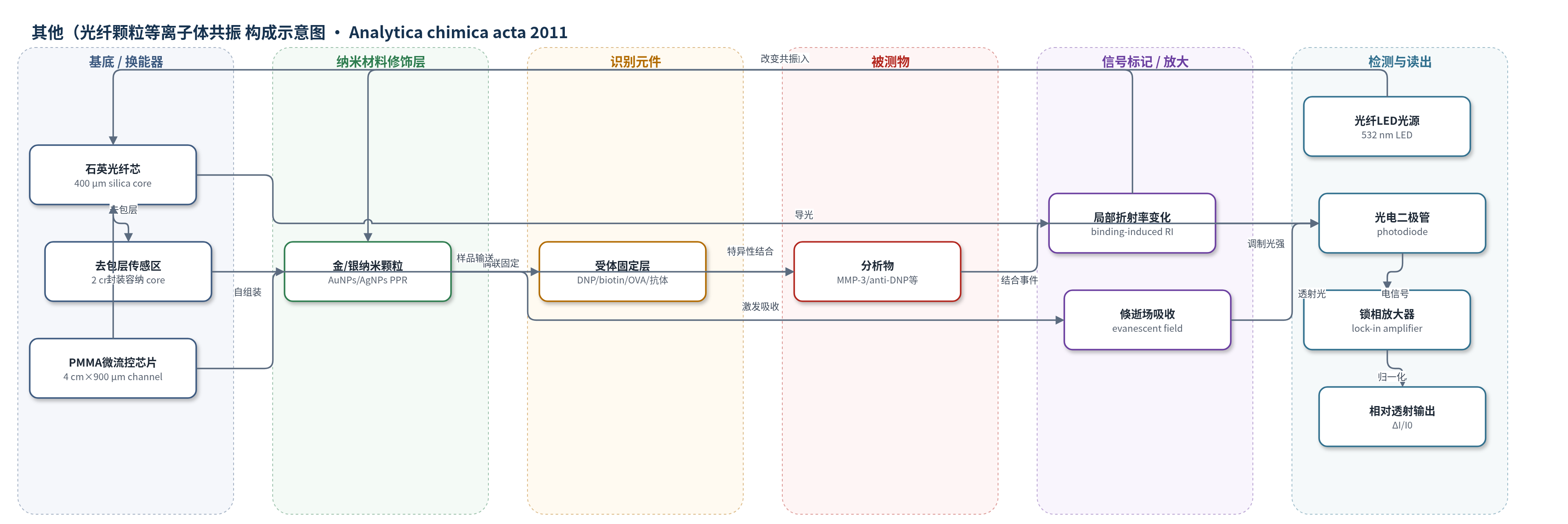
点击图片查看大图 · 依据论文自动绘制
传感器类型
其他(光纤颗粒等离子体共振(FO-PPR)微流控生物传感器)
检测对象
链霉亲和素(streptavidin)、抗生物素抗体(anti-biotin)、抗DNP抗体(anti-DNP)、抗OVA抗体(anti-OVA)、基质金属蛋白酶-3(MMP-3);样品基质:缓冲液及骨关节炎患者滑液(synovial fluid)
检测原理
FO-PPR传感器基于光纤倏逝场吸收。532 nm光在多模光纤中经多次全内反射,在去包层纤芯表面形成倏逝场,激发表面金或银纳米颗粒的颗粒等离子体共振(PPR/LSPR),产生特征吸收。纳米颗粒表面固定DNP、生物素、OVA或抗MMP-3等受体;当分析物如MMP-3、anti-DNP等结合后,纳米颗粒周围局部折射率升高,PPR吸收增强,透射光强降低。系统以ΔI/I0=(I0-IS)/I0为输出,信号随结合量/浓度增加而增大。微流控芯片将传感区缩小至约14 μL,缩短扩散距离并提高界面/体积比,使质量传输更快,从而缩短响应时间并降低检测限。该过程无需标记物或酶催化放大。
检测灵敏度
LOD(原文称DL): 微流控 DL for streptavidin 1.1 × 10⁻¹¹ M、DL for anti-biotin 3.3 × 10⁻⁹ M、DL for anti-DNP 1.2 × 10⁻¹² M;传统液池 DL for streptavidin 9.8 × 10⁻¹¹ M、DL for anti-biotin 8.5 × 10⁻⁸ M、DL for anti-DNP 4.0 × 10⁻¹¹ M;AgNPs anti-biotin DL 2.1 × 10⁻⁹ M、quantification limit 3.0 × 10⁻⁹ M;RIR: 2.9 × 10⁻⁵ RIU(微流控)、7.0 × 10⁻⁵ RIU(传统);r = 0.9996。
效应效果
选择性:DNP芯片对anti-DNP响应随浓度升高,anti-HSA、HSA、BSA无显著变化。重现性:AgNPs芯片检测anti-biotin,3芯片校准r=0.9996,CV为4.0%–21.1%,排除最低浓度后4.0%–11.7%。稳定性:OVA/anti-OVA经3次结合–再生循环,结合CV=3.6%,再生基线CV=2.4%;无受体芯片室温保存至少2个月。实际样品:6例滑液MMP-3中FO-PPR与ELISA配对t检验P=0.8747,无显著差异。微流控使RIR由7.0×10−5 RIU降至2.9×10⁻⁵ RIU,t95由2.72×10^3 s降至2.94×10^2 s,检测限改善约9–33倍,具低成本便携潜力。
传感器的构成
- 基底/换能器:多模塑料包层石英光纤(F-MBC,芯径400 μm、包层430 μm),部分去包层2 cm,作为光波导与倏逝场换能器。
- 微流控芯片:聚甲基丙烯酸甲酯(PMMA)上下板,流道4.0 cm×900 μm×900 μm,自由体积约14 μL,热压合封装,用于样品输送与检测池。
- 纳米材料修饰层:纤芯表面经3-巯基丙基三甲氧基硅烷(MPTMS)自组装固定金纳米颗粒(AuNPs,15.9±2.5 nm)或银纳米颗粒(AgNPs,31.6±8.4 nm),提供颗粒等离子体共振(PPR/LSPR)吸收。
- 识别元件:纳米颗粒表面经半胱胺(cystamine)自组装单分子层(SAM)及EDC/HEPES偶联固定DNP或生物素(biotin),或按先前方法固定OVA或抗MMP-3抗体(anti-MMP-3)。
- 信号标记物:无标记(label-free),结合事件改变纳米颗粒周围局部折射率,引起532 nm透射吸收变化。
- 读出系统:532 nm光纤LED、函数发生器(约1 kHz调制)、锁相放大器、光电二极管和计算机,输出ΔI/I0。
中文摘要
本文报道将光纤颗粒等离子体共振(FO-PPR)生物传感器与微流控芯片集成,以缩短响应时间并改善检测限。聚甲基丙烯酸甲酯(PMMA)微流控芯片的流道尺寸为4.0 cm×900 μm×900 μm,将一段部分去包层的光纤置于流道内,其纤芯表面修饰金或银纳米颗粒,流道自由体积约14 μL。不同折射率蔗糖溶液实验表明,微流控系统中折射率分辨率提高2.4倍。微流控芯片可精确输送生物样品至检测区而不造成稀释。作者选择生物素/链霉亲和素、生物素/抗生物素、DNP/抗DNP、OVA/抗OVA和抗MMP-3/MMP-3等受体–分析物对考察传感性能。结果显示,达到平衡的响应时间由传统液池的数千秒缩短至微流控流池的数百秒,检测限改善约一个数量级。以相对透射响应变化作为输出可减轻对精确光路对准的要求,并获得较好的芯片间测量重现性。
英文摘要
This article reports the integration of the fiber optic-particle plasmon resonance (FO-PPR) biosensor with a microfluidic chip to reduce response time and improve detection limit. The microfluidic chip made of poly(methyl methacrylate) had a flow-channel of dimensions 4.0 cm × 900 μm × 900 μm. A partially unclad optical fiber with gold or silver nanoparticles on the core surface was placed within the flow-channel, where the volume of the flow space was about 14 μL. Results using sucrose solutions of various refractive indexes show that the refractive index resolution improves by 2.4-fold in the microfluidic system. The microfluidic chip is capable of delivering a precise amount of biological samples to the detection area without sample dilution. Several receptor/analyte pairs were chosen to examine the biosensing capability of the integrated platform: biotin/streptavidin, biotin/anti-biotin, DNP/anti-DNP, OVA/anti-OVA, and anti-MMP-3/MMP-3. Results show that the response time to achieve equilibrium can be shortened from several thousand seconds in a conventional liquid cell to several hundred seconds in a microfluidic flow-cell. In addition, the detection limit also improves by about one order of magnitude. Furthermore, the normalization by using the relative change of transmission response as the sensor output alleviate the demand on precise optical alignment, resulting in reasonably good chip-to-chip measurement reproducibility.