组成图示
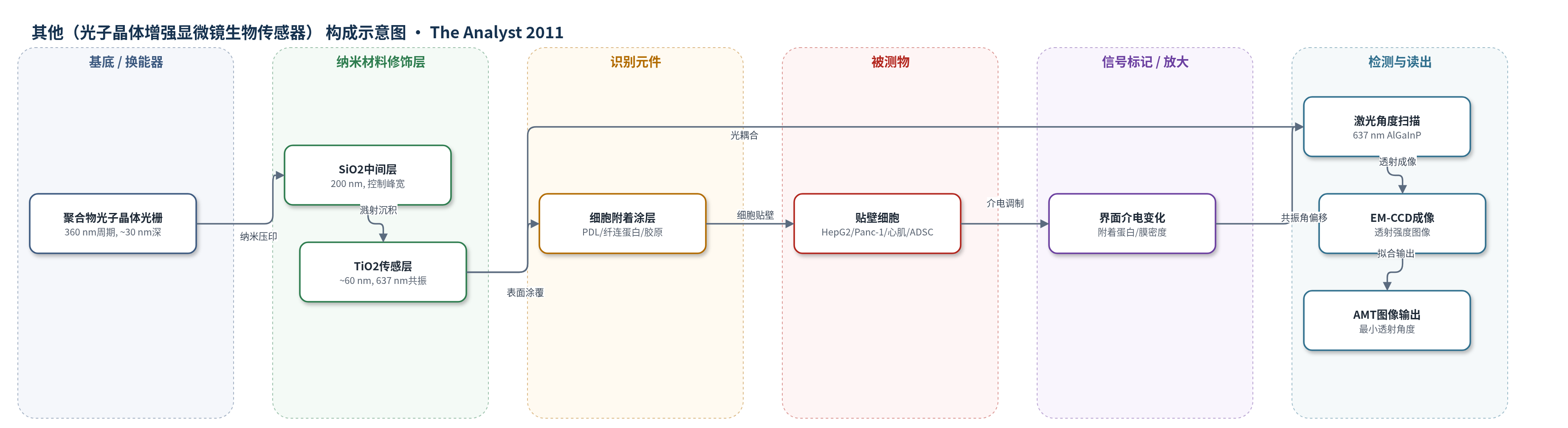
点击图片查看大图 · 依据论文自动绘制
传感器类型
其他(光子晶体增强显微镜生物传感器)
检测对象
细胞附着密度(cell attachment density);样品基质为细胞培养液(DMEM/MEM 含 10% FBS)中的 HepG2/C3、Panc-1、新生大鼠心肌细胞、猪脂肪来源干细胞(ADSC)。
检测原理
PCEM利用聚合物光子晶体光栅作为光学谐振器,637 nm TE偏振激光从下方以0.01°步进扫描入射角。当入射角满足共振耦合条件时,光被高效反射,透射强度出现最小值。细胞通过整合素和丝状伪足贴附到涂层表面后,细胞-传感器界面附近约100 nm倏逝场区域内的介电常数因附着蛋白和膜密度增加而升高,使局部共振耦合角向更低角度偏移。软件对每个像素的透射强度-角度曲线拟合,得到最小透射角度(AMT)图像。附着越强、附着蛋白密度越高,AMT偏移越大;细胞收缩、凋亡或分化导致附着减弱时,AMT偏移减小。系统不依赖HCR、RCA或酶催化沉积等放大策略,信号直接反映界面介电变化。
检测灵敏度
—
效应效果
PCEM可无标记连续监测同一批细胞数天,避免细胞毒性染色。空间分辨率小于1 μm,时间分辨率约1帧/分钟。HepG2/C3细胞1 h培养后平均AMT偏移0.25±0.01°;Panc-1细胞staurosporine处理前AMT偏移0.10°,处理后附着下降。心肌细胞铺展态较圆形态AMT偏移显著增大(p=0.0133和p<0.0001),收缩细胞附着更强(p=0.0133);ADSC神经诱导后附着显著降低(p=0.0007)。由于检测角度偏移而非强度,对瞬时颗粒不敏感;细胞脱离后几乎不留痕迹。相比SPRi,其传播长度可小于5 μm,分辨率更高。
传感器的构成
- 基底/换能器:塑料基底上的聚合物光子晶体光栅(PC grating,周期360 nm、深度约30 nm),作为低折射率亚波长谐振器并限制横向光传播
- 中间层:200 nm SiO2蒸发层,控制共振峰宽度并形成平缓曲面
- 传感表层:约60 nm TiO2射频溅射层,设定637 nm水环境约4°共振条件
- 载体/封装:1×3 in^2显微镜载玻片与六孔无底微孔板,用UV固化胶AC R260-A1键合
- 识别/附着层:聚-L-赖氨酸PDL(100 ng/mL)或纤维连接蛋白/胶原(5 mg/mL fibronectin + 20 mg/mL collagen)或胶原(25 mg/mL),提供细胞贴壁配体
- 识别元件:细胞表面整合素(integrin)与丝状伪足(filopodia),介导细胞与涂层/传感器界面结合
- 信号标记物:无外加标记,细胞附着蛋白与膜界面密度作为内源介电信号源
- 读出系统:637 nm AlGaInP激光、旋转镜、10×/20×物镜、EM-CCD相机与AMT拟合软件,输出最小透射角度图像
中文摘要
本文提出光子晶体增强显微镜(PCEM)作为一种无标记生物传感器成像技术,用于测量细胞表面附着及附着调制。该方法将光子晶体光学谐振器表面集成到常规微孔板孔中,并采用显微镜检测系统测量细胞-传感器界面介电常数局部变化引起的共振耦合条件偏移。研究在四个模型体系中验证:HepG2/C3肝癌细胞用于表征生长与运动背景下的细胞黏附;Panc-1胰腺癌细胞用于验证staurosporine诱导凋亡时细胞附着密度下降;原代新生心肌细胞用于确认整合素介导信号对生长和发育的影响,其中圆形细胞附着密度降低,铺展细胞附着强度与收缩性增强;猪脂肪来源干细胞用于监测强制分化过程中的形态变化与细胞外基质重塑。上述实验均无需潜在细胞毒性标记,即可对同一批细胞进行数天连续观察。
英文摘要
We introduce photonic crystal enhanced microscopy (PCEM) as a label-free biosensor imaging technique capable of measuring cell surface attachment and attachment modulation. The approach uses a photonic crystal optical resonator surface incorporated into conventional microplate wells and a microscope-based detection instrument that measures shifts in the resonant coupling conditions caused by localized changes in dielectric permittivity at the cell-sensor interface. Four model systems are demonstrated for studying cancer cells, primary cardiac muscle cells, and stem cells. First, HepG2/C3 hepatic carcinoma cells were cultured and observed via PCEM in order to characterize cell adhesion in the context of growth and locomotion. Second, Panc-1 pancreatic cancer cells were used to verify that cell attachment density decreases in response to staurosporine, a drug that induces apoptosis. Third, we used PCEM to confirm the influence of integrin-mediated signaling on primary neonatal cardiomyocyte growth and development. Rounded cardiomyocytes consistently showed decreased cell attachment density as recorded via PCEM, while spreading cells exhibited greater attachment strength as well as increased contractility. Finally, PCEM was used to monitor the morphological changes and extracellular matrix remodeling of porcine adipose-derived stem cells subjected to a forced differentiation protocol. Each of these experiments yielded information regarding cell attachment density without the use of potentially cytotoxic labels, enabling study of the same cells for up to several days.