组成图示
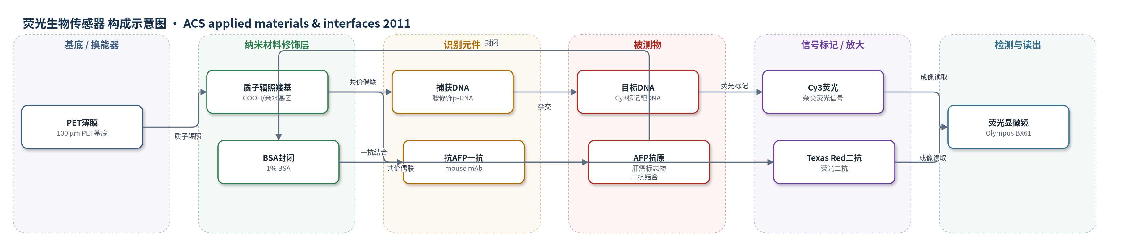
点击图片查看大图 · 依据论文自动绘制
传感器类型
荧光生物传感器
检测对象
炭疽毒素目标DNA(anthrax toxin target DNA,杂交缓冲液)、α-胎蛋白(α-fetoprotein, AFP,抗原溶液)
检测原理
PET表面经200 keV质子辐照产生羧基,经EDC/NHS活化后与生物分子氨基共价偶联,实现图案化固定。DNA通道中,胺修饰捕获DNA固定于辐照区,与Cy3标记目标DNA互补杂交,杂交量随目标DNA浓度增加而增加,Cy3荧光强度随之增强。免疫通道中,AFP抗原固定于辐照区并经BSA封闭,抗AFP一抗与抗原结合,Texas Red标记二抗再结合一抗,形成抗原-一抗-二抗复合物;抗原浓度越高,结合的二抗越多,Texas Red荧光越强。最终用荧光显微镜成像,以微图案平均荧光强度反映目标DNA或AFP浓度。
检测灵敏度
LOD: 4 ng/mL (target DNA, 5×10^14 ions/cm2);LOD: 16 ng/mL (target AFP, 5×10^14 ions/cm2)
效应效果
该PET荧光传感平台在5×10^14 ions/cm2辐照下表现优于1×10^14 ions/cm2,因表面羧基含量更高,固定生物分子量更大,荧光信号更强。选择性方面,非互补目标DNA不产生杂交信号;无EDC/NHS时生物分子不能共价固定,无荧光信号;预封闭二抗实验表明免疫识别具有特异性。目标DNA和AFP的最低可检测浓度分别为4 ng/mL和16 ng/mL。作者认为离子辐照功能化PET是制备柔性聚合物基底生物传感器的有前景策略,可用于炭疽毒素和肝癌标志物检测。文中未报告RSD、稳定性、实际样品回收率或与ELISA/qPCR的定量对比。
传感器的构成
- 基底/换能器:PET薄膜(poly(ethylene terephthalate, PET),100 μm,柔性聚合物基底,经200 keV H+离子辐照生成表面羧基,作为生物分子固定与荧光成像基底
- 表面功能化层:质子辐照诱导羧基(COOH)/羟基(OH)等亲水基团,提供共价偶联位点,并经掩模形成图案化区域
- 偶联试剂层:EDC/NHS(1-ethyl-3-(3-dimethylaminopropyl) carbodiimide hydrochloride / N-hydroxysuccinimide),活化PET表面羧基并与生物分子氨基形成酰胺键
- 识别元件(DNA通道):胺修饰炭疽毒素捕获DNA(amine-modified capture DNA/probe DNA),共价固定于辐照区,用于捕获目标DNA
- 抗原固定层:AFP抗原(α-fetoprotein, AFP)固定于辐照区,作为肝癌标志物抗原展示位点
- 识别元件(免疫通道):抗AFP一抗(mouse monoclonal anti-AFP antibody),与固定AFP抗原特异性结合
- 封闭剂:1% BSA(bovine serum albumin),封闭残余NHS酯和非特异结合位点
- 信号标记物:Cy3标记目标DNA(Cy3-labeled target DNA)用于DNA杂交荧光信号;Texas Red标记小鼠IgG二抗(Texas Red-labeled secondary antibody)用于免疫荧光信号
- 检测读出:荧光显微镜(Olympus BX61)与ImageJ分析,读取微图案平均荧光强度
中文摘要
本研究通过不同质子束通量选择性辐照聚对苯二甲酸乙二醇酯(PET)薄膜表面,在辐照区域生成羧基,并对功能化 PET 表面的润湿性、化学结构和化学组成进行表征。结果表明,辐照区成功生成羧基,且其相对含量随通量变化。随后,将炭疽毒素探针 DNA 选择性固定于辐照区,Cy3 标记的目标 DNA 与固定探针 DNA 杂交;同时,将肝癌特异性 α-胎蛋白(AFP)抗原选择性固定于辐照区,并采用间接免疫法,以 AFP 特异性一抗和 Texas Red 标记二抗检测目标抗原。结果显示,在功能化 PET 表面成功形成清晰生物分子微图案,且微图案荧光强度主要取决于与探针 DNA 杂交的目标 DNA 浓度以及固定于 PET 薄膜上的目标 AFP 抗原浓度。在通量为 5×10^14 ions/cm^2 制备的 PET 薄膜上,目标 DNA 和目标 AFP 抗原的最低可检测浓度分别为 4 ng/mL 和 16 ng/mL。
英文摘要
The surface of a poly(ethylene terephthalate) (PET) film was selectively irradiated with proton beams at various fluences to generate carboxylic acid groups on the surface; the resulting functionalized PET surface was then characterized in terms of its wettability, chemical structure, and chemical composition. The results revealed that (i) carboxylic acid groups were successfully generated in the irradiated regions of the PET surface, and (ii) their relative amounts were dependent on the fluence. A capture biomolecule, anthrax toxin probe DNA, was selectively immobilized on the irradiated regions on the PET surface. Cy3-labeled DNA as a target biomolecule was then hybridized with the probe DNA immobilized on the PET surface. Liver-cancer-specific α-fetoprotein (AFP) antigen, as a target biomolecule, was also selectively immobilized on the irradiated regions on the PET surface. Texas Red-labeled secondary antibody was then reacted with an AFP-specific primary antibody prebound to the AFP antigen on the PET surface for the detection of the target antigen, using an indirect immunoassay method. The results revealed that (i) well-defined micropatterns of biomolecules were successfully formed on the functionalized PET surfaces and (ii) the fluorescence intensity of the micropatterns was dependent mainly on the concentrations of the target DNA hybridized to the probe DNA and the target AFP antigen immobilized on the PET films. The lowest detectable concentrations of the target DNA and target AFP antigen in this study were determined to be 4 and 16 ng/mL, respectively, with the PET film prepared at a fluence of 5 × 10(14) ions/cm(2).