组成图示
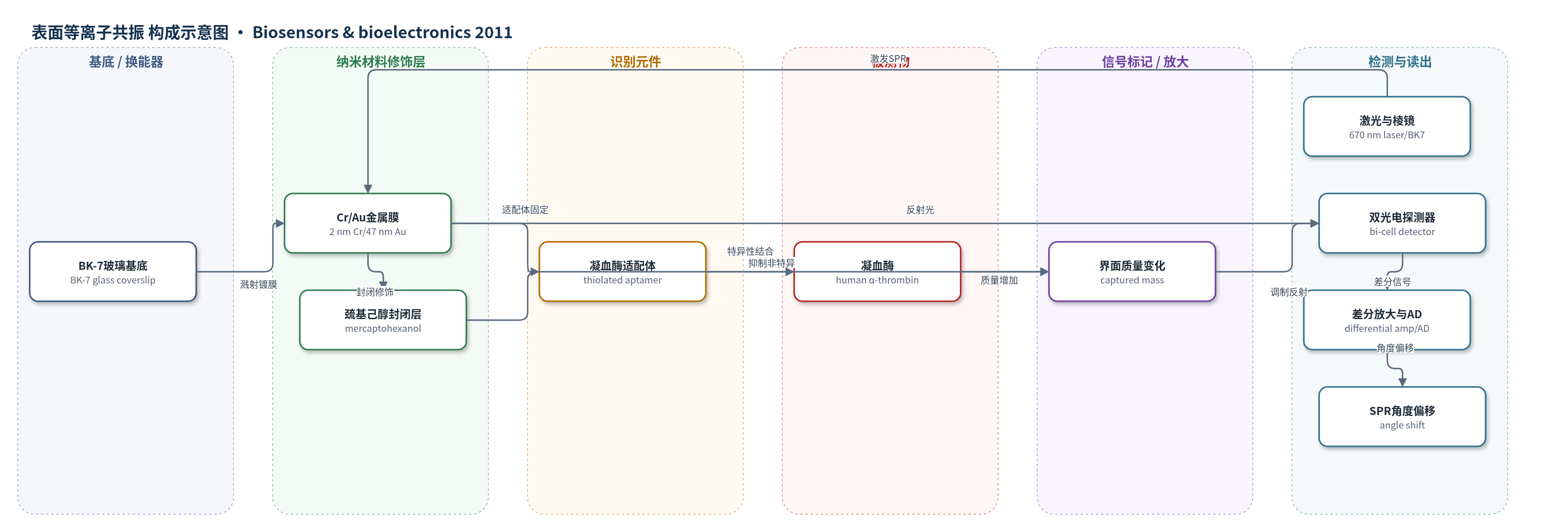
点击图片查看大图 · 依据论文自动绘制
传感器类型
表面等离子共振(SPR)生物传感器
检测对象
凝血酶(thrombin,human α-thrombin);样品基质:Tris–EDTA 缓冲液、10% 胎牛血清(FBS)加标血清样品。
检测原理
该传感器基于 Kretschmann 构型的双细胞 SPR。670 nm 激光经 BK7 棱镜激发金膜表面等离子体,反射光在共振角处出现暗线。凝血酶适配体通过 Au–S 键固定于金表面,6-巯基-1-己醇封闭层降低非特异吸附。样品流过时,凝血酶与适配体结合,传感界面捕获质量增加,局部折射率改变,使共振角偏移。双光电探测器采集偏移前后反射光强,差分放大器计算 (A−B)/(A+B),该比值与共振角变化成正比,从而实时、无标记地反映凝血酶浓度。血清中非特异蛋白吸附和流速会引起背景漂移,需稀释或参考通道校正。
检测灵敏度
LOD: 25 nM (Tris–EDTA buffer);LOD: 50 nM (serum spiked with thrombin);线性范围: 50 nM–200 nM;R^2 = 0.99 (buffer linearity);R^2 = 0.9992 (intra-day);R^2 = 0.9892 (inter-day)
效应效果
检测对凝血酶适配体和凝血酶均特异:凝血因子 X、错配适配体和仅封闭层无显著 SPR 偏移;200 nM 凝血酶原可被检出,但 ≤100 nM 时未检出,提示临床样品中凝血酶原干扰有限。缓冲液中 50、100、200 nM 凝血酶的日内 CV 分别为 12.21%、12.14%、20.13%,日间 CV 分别为 12.43%、13.09%、18.6%。10% FBS 加标血清中 LOD 为 50 nM,线性范围 50–200 nM,但存在持续低水平背景漂移;BSA 预封闭或 0.05% Tween 20 不能消除漂移。作者认为该平台实时、无标记、成本低于商用仪器,可适配临床检测。
传感器的构成
- 基底:BK-7 玻璃盖玻片(BK-7 glass coverslips),承载金属膜并作为 SPR 基底
- 金属换能层:2 nm Cr/47 nm Au 溅射层,形成表面等离子共振界面
- 识别元件:巯基化 DNA 凝血酶适配体(thiolated thrombin aptamer,15-mer,3′-ThioMC3-D),经 Au–S 键固定并特异性结合凝血酶 exosite 1
- 封闭层:6-巯基-1-己醇(mercaptohexanol,SH-(CH2)6-OH)自组装单分子层,封闭金表面以降低非特异吸附
- 流路单元:4 μL 注射流池(Teflon flow cell,5 mm² 传感面积),使 Tris–EDTA 或 10% FBS 样品以 10 μL/min 流过传感面
- 光学读出:670 nm 二极管激光、BK7 半圆柱棱镜、双光电探测器(bi-cell photodetector)、差分放大器与 AD 采集,输出 SPR 角度偏移
中文摘要
丝氨酸蛋白酶凝血因子凝血酶主要参与止血,也与动脉粥样硬化、血栓栓塞性疾病、癌症和炎症疾病相关。由于缺乏特异性探针且凝血酶浓度低,血浆中凝血酶等凝血蛋白的直接测定具有挑战;样品中高蛋白浓度还会造成高背景。本研究采用双细胞表面等离子共振(SPR)光谱仪,将凝血酶适配体固定于低体积流池传感表面,对样品中凝血酶进行实时、无标记检测。在Tris–EDTA缓冲液中,凝血酶检出限(LOD)为25 nM;50 nM凝血酶日内和日间检测的变异系数(CV)分别为12.2%和12.4%。该检测对凝血酶适配体和凝血酶均具有特异性。在加标血清样品中,LOD为50 nM,线性检测范围为50–200 nM,但血清样品存在持续的低水平背景漂移。作者评估了传感表面非特异蛋白吸附和样品流速的贡献,并探讨了降低背景漂移的策略。结果表明,双细胞SPR平台结合适配体捕获探针可作为高灵敏度实时无标记生物传感器,用于血浆样品中凝血因子的检测。
英文摘要
The serine protease coagulation factor thrombin functions primarily in hemostasis, but is also involved in atherosclerosis, thromboembolic disease, cancer and inflammatory disease. Direct measurement of coagulation proteins including thrombin in plasma samples poses a significant challenge because of lack of specific probes and low thrombin concentrations. In addition, high plasma protein concentrations in samples can result in high backgrounds. These challenges were overcome using a bi-cell surface plasmon resonance (SPR) spectrometer with an immobilized thrombin aptamer to measure thrombin in samples passed through a low volume flow cell. For thrombin in Tris-EDTA buffer, the limit of detection (LOD) was 25 nM. Coefficient of variation (CV) for detection of 50 nM was 12.2% and 12.4% for intra and inter-day measurements respectively. This detection was specific for both thrombin aptamer and for thrombin. Using serum samples spiked with thrombin, the LOD was 50 nM with a linear range of detection from 50 nM to 200 nM. However use of serum samples was associated with consistent, low-level background drift. The contributions of nonspecific protein absorption onto the sensor surface and sample flow speed were assessed, and strategies to reduce this background drift were explored. We conclude that the bi-cell SPR platform with an aptamer capture probe can be employed as a highly sensitive real-time, label-free biosensor for the detection of coagulation factors in plasma samples.