组成图示
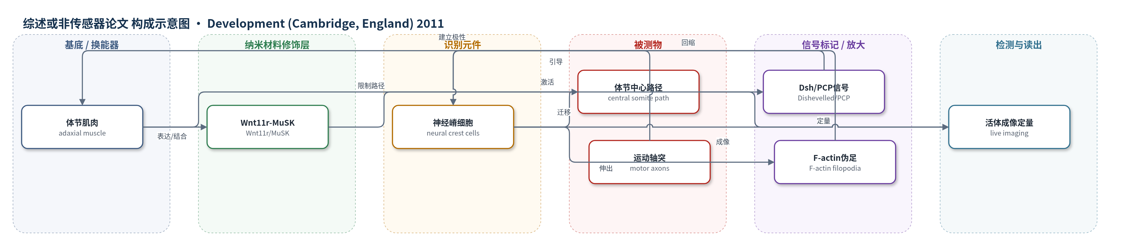
点击图片查看大图 · 依据论文自动绘制
传感器类型
综述或非传感器论文
检测对象
不适用(整体非传感器论文);文中F-actin荧光探针的检测对象为F-actin(肌动蛋白纤维),样品基质为斑马鱼胚胎神经嵴细胞。
检测原理
本文非传感检测论文,未涉及换能器、识别元件或信号放大策略。其生物学机制为:体节肌肉细胞表达MuSK,接受分泌糖蛋白Wnt11r信号后激活Dishevelled(Dsh)依赖的非经典Wnt/平面细胞极性(PCP)通路,在肌肉细胞内建立前后轴中心区,并通过细胞外基质(ECM)组分(如CSPG、Tenascin C)限制神经嵴细胞进入体节中心路径。MuSK缺失或Wnt11r缺失时,神经嵴细胞仍能初始进入分节路径,但随后向体节侧方扩散;F-actin生物传感器显示其前缘丝状伪足不能有效回缩,导致迁移轨迹变宽。运动轴突可引导神经嵴,但并非分节迁移所必需。
检测灵敏度
原文未报告LOD、线性范围、灵敏度斜率或相关系数。
效应效果
论文未评价传感器选择性、抗干扰、稳定性、重现性或加标回收率,也未与ELISA/HPLC/qPCR对比。生物学表型:plexinA3突变中82%(n=48半体节)神经嵴跟随异位运动轴突;musk突变中60%(n=80)运动轴突偏离中心,53/80神经嵴侵入更宽区域;wnt11r突变中25%(n=335)运动轴突缺陷,20%(n=120)神经嵴偏离中心,10%独立轨迹;肌肉表达Myc-Dsh-DEP+约46%(n=55)重现缺陷。运动神经元消融后,野生型神经嵴宽度/体节宽度约0.25,musk和wnt11r分别升高近2倍和大于2倍。E10.5小鼠Musk敲除(n=5)神经嵴不再限于前体节。作者主张该通路进化保守地维持分节迁移。
传感器的构成
- 基底/换能器:不适用(非传感器论文;活体成像对象为斑马鱼/小鼠胚胎)
- 纳米材料修饰层:不适用(非传感器论文;未报道纳米材料修饰)
- 荧光探针:Lifeact-GFP/mCherry-UtrCH,标记总/稳定F-actin
中文摘要
躯干神经嵴细胞从背侧神经管以连续片状脱离,但在穿过体节区域前转变为分节排列的迁移流。这些神经嵴细胞流与寻找路径的脊髓运动轴突共享分节轨迹,提示二者相互作用可能影响神经嵴迁移。本文显示,在斑马鱼胚胎中,神经嵴细胞与运动轴突的迁移在时间上同步,并空间限制于体节中心;但运动轴突并非分节神经嵴迁移所必需。相反,肌肉特异性受体激酶(MuSK)及其推测配体Wnt11r对将神经嵴细胞限制在每个体节中心至关重要。此外,阻断体节肌肉细胞中的平面细胞极性(PCP)信号也导致非分节神经嵴迁移。利用F-actin生物传感器发现,缺乏MuSK时神经嵴细胞不能回缩无效的前缘突起,从而出现非分节迁移。最后,MuSK敲除小鼠表现出相似的神经嵴迁移缺陷,提示MuSK在神经嵴迁移中具有新的、进化保守的作用。作者提出Wnt11r-MuSK依赖的PCP样通路限制神经嵴细胞沿分节路径迁移。
英文摘要
Trunk neural crest cells delaminate from the dorsal neural tube as an uninterrupted sheet; however, they convert into segmentally organized streams before migrating through the somitic territory. These neural crest cell streams join the segmental trajectories of pathfinding spinal motor axons, suggesting that interactions between these two cell types might be important for neural crest cell migration. Here, we show that in the zebrafish embryo migration of both neural crest cells and motor axons is temporally synchronized and spatially restricted to the center of the somite, but that motor axons are dispensable for segmental neural crest cell migration. Instead, we find that muscle-specific receptor kinase (MuSK) and its putative ligand Wnt11r are crucial for restricting neural crest cell migration to the center of each somite. Moreover, we find that blocking planar cell polarity (PCP) signaling in somitic muscle cells also results in non-segmental neural crest cell migration. Using an F-actin biosensor we show that in the absence of MuSK neural crest cells fail to retract non-productive leading edges, resulting in non-segmental migration. Finally, we show that MuSK knockout mice display similar neural crest cell migration defects, suggesting a novel, evolutionarily conserved role for MuSK in neural crest migration. We propose that a Wnt11r-MuSK dependent, PCP-like pathway restricts neural crest cells to their segmental path.