组成图示
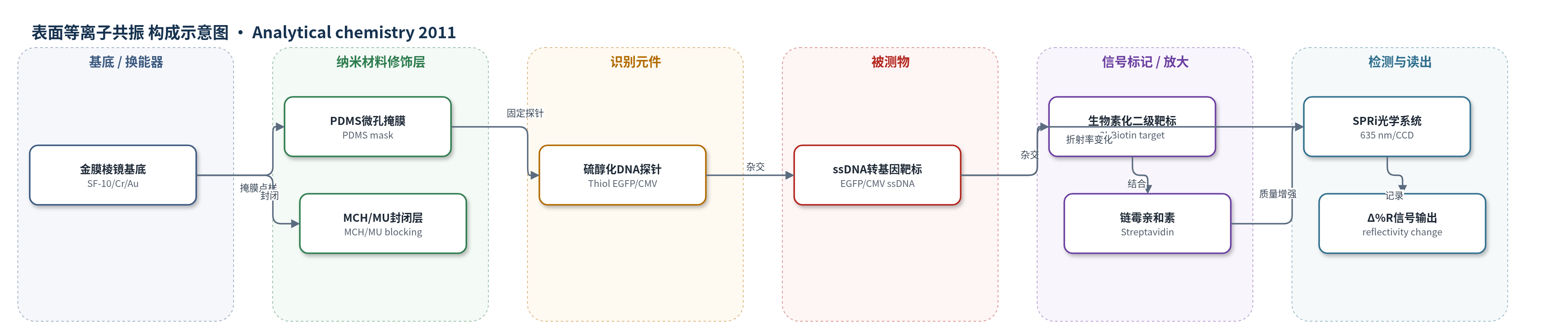
点击图片查看大图 · 依据论文自动绘制
传感器类型
表面等离子共振(SPR)生物传感器
检测对象
转基因DNA标记序列(CMV promoter、EGFP gene);样品基质:HEK细胞基因组DNA的PCR单链扩增产物及合成寡核苷酸
检测原理
传感器在金膜表面固定硫醇化DNA探针,形成多序列识别阵列。PCR双链扩增产物经链霉亲和素磁珠碱解离后,释放单链转基因靶标;靶标与互补探针杂交,使金膜界面质量与折射率增加。SPRi以635 nm p偏振光照射金膜,界面折射率变化引起反射率变化,CCD实时记录Δ%R。靶标浓度越高,结合量越大,Δ%R越大,直至探针饱和。为增强信号,3′-生物素化二级靶标与已结合靶标杂交,再结合链霉亲和素,通过生物素–链霉亲和素高亲和作用实现原位质量放大。
检测灵敏度
LOD: 12 nM(EGFP1)、0.2 nM(EGFP2)、3.5 nM(CMV);线性范围: 1–500 nM(校准范围),EGFP1 线性范围达 250 nM,EGFP2 与 CMV 约 60 nM 饱和;灵敏度斜率: EGFP2 在 0–50 nM 区间斜率最高;最小可记录信号: 0.06 Δ%R(3×SD,SD = 0.02 Δ%R)
效应效果
系统选择性高,各靶标仅与互补探针结合,非互补探针和参考点无显著信号。校准点三次重复CV%均<10%。SPRi检出限较压电传感器低约1个数量级:EGFP2为0.2 nM(压电50 nM),CMV为3.5 nM(压电25 nM),EGFP1为12 nM(压电50 nM)。PCR样品中,HEK-293阴性平均0.009±0.02 Δ%R,EGFP2为0.34±0.02 Δ%R,CMV为0.07±0.02 Δ%R;qPCR测得HEK-GFP为132.01±9.88拷贝/0.5 ng DNA,接近理论133拷贝。原位放大仅EGFP2信号显著增加。作者认为可用于基因兴奋剂高通量无标记检测。
传感器的构成
- 基底/换能器:SF-10 玻璃棱镜,5 nm Cr 底层与 50 nm Au 层,用于 SPRi 光学换能
- 点样掩膜:PDMS 微孔掩膜(184 Silicone Elastomer),用于分区固定探针(制备后移除)
- 识别元件:硫醇化 ssDNA 探针(Thiol EGFP1、Thiol EGFP2、Thiol CMV),通过 Au–S 自组装固定并识别互补转基因 DNA
- 封闭层:1 μM MCH 与 1 μM MU 混合硫醇,封闭金表面非特异结合位点
- 样品预处理:生物素化 PCR 引物扩增产物与 SA-PMPs 链霉亲和素磁珠,碱解离后释放 ssDNA 靶标
- 信号放大:3′-生物素化二级靶标(EGFP1_RFBiot30、EGFP2_RFBiot30、CMV_RFBiot30)与已结合靶标杂交
- 信号放大:链霉亲和素(Streptavidin)结合生物素,增加界面质量以增强 SPRi 信号
- 结合液/读出:BS(300 mM NaCl、20 mM Na2HPO4、0.1 mM EDTA、pH 7.4)与 SPRi-Lab+ 635 nm 光路,CCD 记录 Δ%R
中文摘要
本文以表面等离子共振成像(SPRi)为换能原理,开发用于人细胞系中转基因检测的光学生物传感方法。研究目标是建立一种多分析物、无标记、实时检测转基因事件标记 DNA 序列的策略。该方法通过识别穿梭载体骨架上用于人胚胎肾(HEK)细胞转染的特定标记序列来判定转基因事件,文中选定了巨细胞病毒启动子(CMV)和增强型绿色荧光蛋白(EGFP)基因序列作为靶标。论文讨论了探针效率、二级结构对传感器表面生物识别反应的影响,并优化了 PCR 样品预处理流程,使双链扩增产物能够与芯片探针杂交;同时提出通过原位质量增强提高 SPRi 信号。实时 PCR(qPCR)作为参考方法用于检测人 HEK 细胞中的标记序列。作者认为该系统有望应用于反兴奋剂领域中的基因兴奋剂检测。
英文摘要
Surface plasmon resonance imaging (SPRi) was used as the transduction principle for the development of optical-based sensing for transgenes detection in human cell lines. The objective was to develop a multianalyte, label-free, and real-time approach for DNA sequences that are identified as markers of transgenosis events. The strategy exploits SPRi sensing to detect the transgenic event by targeting selected marker sequences, which are present on shuttle vector backbone used to carry out the transfection of human embryonic kidney (HEK) cell lines. Here, we identified DNA sequences belonging to the Cytomegalovirus promoter and the Enhanced Green Fluorescent Protein gene. System development is discussed in terms of probe efficiency and influence of secondary structures on biorecognition reaction on sensor; moreover, optimization of PCR samples pretreatment was carried out to allow hybridization on biosensor, together with an approach to increase SPRi signals by in situ mass enhancement. Real-time PCR was also employed as reference technique for marker sequences detection on human HEK cells. We can foresee that the developed system may have potential applications in the field of antidoping research focused on the so-called gene doping.