组成图示
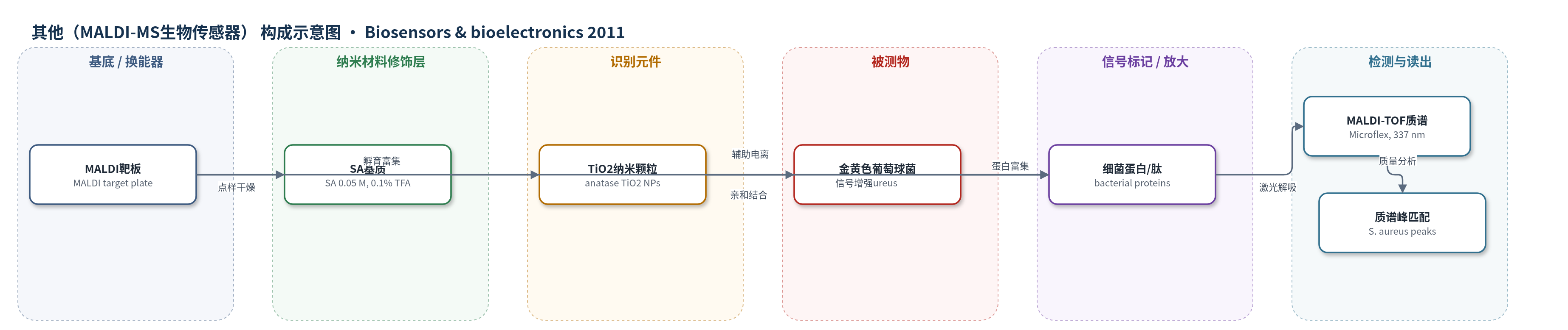
点击图片查看大图 · 依据论文自动绘制
传感器类型
其他(MALDI-MS生物传感器)
检测对象
金黄色葡萄球菌(Staphylococcus aureus, S. aureus);样品基质:空气、人体皮肤表面拭子、人体鼻腔拭子(无菌水悬浮液)
检测原理
样品中的金黄色葡萄球菌与锐钛矿型 TiO2 纳米颗粒发生亲和相互作用,TiO2 NPs 作为亲和探针选择性富集细菌蛋白/肽并增强 MALDI 离子化效率。随后加入 SA 基质,基质吸收 337 nm 激光能量,使细菌蛋白/肽解吸电离。MALDI-TOF 按质荷比分离离子,产生特征质谱峰,并与标准 S. aureus 峰匹配以判断目标菌存在。TiO2 NPs 的高比表面提高捕获与信号放大,但浓度过高会因杀菌作用导致信号淬灭,因此采用 1 mg/mL。信号强度随细菌/蛋白丰度增加而增强,但原文未给出定量线性关系。
检测灵敏度
原文未报告本方法 LOD、线性范围、灵敏度斜率或相关系数。
效应效果
直接 MALDI-MS 无 TiO2 时,空气和皮肤样品未检出信号,鼻腔样品仅见少量峰;TiO2 NPs 辅助后,空气和鼻腔样品出现与标准 S. aureus 匹配的特征峰,皮肤样品出现少数匹配峰,说明 TiO2 能选择性增强细菌蛋白并抑制皮肤/鼻腔蛋白干扰。培养计数显示空气、皮肤、鼻腔样品细菌数分别为 9×10^2、7×10^2、4.6×10^2 cfu/mL;培养后菌落刮取 MALDI-MS 进一步确认。方法无需培养和前处理,作者认为其简单、快速、灵敏,适用于临床与环境样品中 S. aureus 检测;未报告 RSD、回收率、稳定性及与 ELISA/PCR 的定量对比。
传感器的构成
- 基底/样品靶板:MALDI target plate,承载样品与基质,供激光解吸
- 亲和/信号增强纳米材料:anatase TiO2 nanoparticles (NPs, 10–20 nm, 1 mg/mL),选择性富集细菌蛋白并增强 MALDI 信号
- 识别/捕获元件:未修饰 anatase TiO2 NPs 作为亲和探针,与细菌细胞/蛋白相互作用
- 基质:3,5-dimethoxy-4-hydroxycinnamic acid (SA, 0.05 M in acetonitrile:water 3:1 v/v, 0.1% TFA),吸收激光并辅助解吸电离
- 被测物/样品:空气、皮肤表面、鼻腔拭子中的 Staphylococcus aureus (S. aureus) 及其蛋白/肽
- 换能/读出仪器:MALDI-TOF mass spectrometer (Bruker Microflex, N2 laser 337 nm, +20 kV, linear mode),将蛋白/肽质量信号转为质谱峰
中文摘要
本文首次将质谱作为生物传感器,用于检测临床重要病原菌金黄色葡萄球菌(Staphylococcus aureus, S. aureus)。作者采用 TiO2 纳米颗粒(NPs)辅助的 MALDI-MS,对空气、鼻腔和皮肤样品进行无培养、快速、直接分析。当金黄色葡萄球菌在个体鼻腔中占优势时,可能在手术或伤口情况下导致伤口感染。研究表明,即使细菌浓度很低,也可从混合菌群中直接检测出该菌,无需培养或其他样品前处理。该方法简单、快速、直接且灵敏,可广泛应用于临床和环境样品中该致命病原菌的检测。
英文摘要
For the first time, we have demonstrated the use of mass spectrometry as a biosensor for detecting a clinically important bacterium: Staphylococcus aureus in air, nasal passage and skin samples using culture-free, rapid, direct analysis via TiO(2) nanoparticles (NPs) assisted MALDI-MS. When this bacterium is predominating, the nasal passage of an individual is observed to lead to wound infections especially when the individual has a surgery or some wounds. This study indicates that even at very low concentrations of an individual bacterium can be directly detected from a mixture of bacteria using the MALDI-MS analysis without the requirement of any culturing steps or any other sample pretreatment techniques. The current approach is extremely simple, rapid, straightforward and sensitive which could be widely applied for the detection of this deadly pathogen in clinical as well as environmental samples.