组成图示
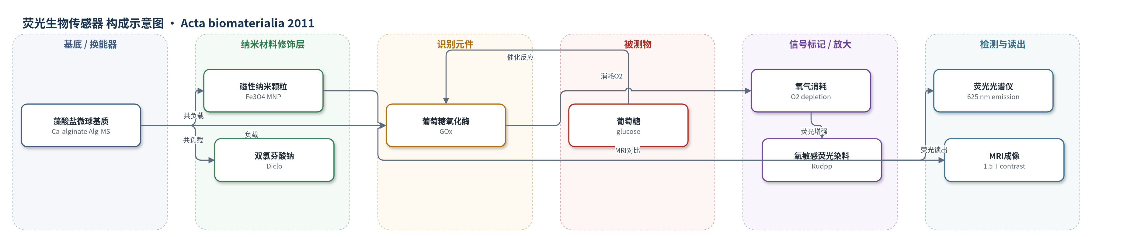
点击图片查看大图 · 依据论文自动绘制
传感器类型
荧光生物传感器
检测对象
葡萄糖(glucose);样品基质:体外葡萄糖溶液(0–50 mM,PBS/水溶液)
检测原理
该传感器以藻酸盐微球为可植入基质,GOx 作为识别元件催化葡萄糖氧化:葡萄糖与溶解氧在 GOx 作用下生成丙酮酸和过氧化氢,使微球内 O2 浓度下降。Rudpp 为氧敏感荧光染料,其荧光可被 O2 动态淬灭;葡萄糖浓度升高时,O2 消耗增加,Rudpp 荧光淬灭减弱,625 nm 发射增强。通过荧光强度比 I/I0 对葡萄糖浓度作图,在 0–10 mM 范围内获得线性响应,回归系数 0.974,响应时间约 3–4 min。该机制不依赖电极或 HCR/RCA 等核酸放大,而是依靠酶催化氧消耗实现光学换能;微球尺寸、GOx 浓度和 O2 扩散速率决定灵敏度与线性范围。Fe3O4 MNP 不直接参与荧光传感,主要提供 MRI 对比。
检测灵敏度
LOD: 0.5 mM;线性范围: 0–10 mM;灵敏度: 3.349% mM^-1;回归系数: 0.974
效应效果
葡萄糖传感 0–10 mM 线性良好,回归系数 0.974,灵敏度 3.349% mM^-1,较 0.52% mM^-1 提高约 6 倍;Rudpp 泄漏 <1%。GOx 活性 2.3×10^5 AU μg^-1 s^-1,1 周降 47%,后 3 周稳定。MNP 使 60/10 μm 微球 24 h 突释率由 17.35/23.68% 增至 28.89/35.87%;85% 释放时间由 30/20 天缩至 21/14 天。VSM 超顺磁性,磁矩 2.09/1.368 emu g^-1,无磁滞;MRI 对比随 MNP 增强。细胞活力 >80%,部分 >95%。可用于植入监测、抗炎与可视化。
传感器的构成
- 微球基质:低粘度藻酸钠(sodium alginate)经氯化钙(CaCl2)交联形成钙藻酸盐微球(Alg-MS),作为可植入载体与扩散通道
- 纳米功能模块:柠檬酸包覆氧化铁磁性纳米颗粒(Fe3O4 MNP),作为 MRI 对比剂并改变微球孔道与药物释放
- 识别元件:葡萄糖氧化酶(GOx,Aspergillus niger)负载于微球内,催化葡萄糖氧化并消耗溶解氧
- 信号标记物:三(4,7-二苯基-1,10-菲啰啉)氯化钌(II)(Rudpp)氧敏感荧光染料,碱性条件下静电沉淀于藻酸盐基质,O2 淬灭其荧光
- 药物模块:双氯芬酸钠(Diclo)负载于微球内,作为非甾体抗炎药抑制植入相关炎症
中文摘要
本研究开发并表征了一种可植入的多功能藻酸盐微球系统,集生物传感、药物递送和磁共振成像(MRI)于一体。系统以葡萄糖氧化酶(GOx)和氧敏感荧光染料三(4,7-二苯基-1,10-菲啰啉)氯化钌(II)(Rudpp)构成葡萄糖生物传感器,以双氯芬酸钠(Diclo)作为抗炎药,以柠檬酸包覆氧化铁磁性纳米颗粒(Fe3O4 MNP)作为 MRI 对比剂。MNP 通过共沉淀法合成,并与 GOx、Diclo 一起经商用滴液发生器包埋于藻酸盐微球中。光学显微镜、SEM、TEM、XRD、VSM 和 MRI 表征表明,MNP 粒径为 5–15 nm,最终微球粒径为 10–60 μm。葡萄糖传感在 0–10 mM 范围内呈良好线性,回归系数为 0.974,响应时间约 4 min。体外释放显示,MNP 负载使 60 和 10 μm 微球的突释率提高 11–12%,但 85% 药物释放持续时间分别缩短 7 和 6 天。VSM 证实超顺磁性,MRI 显示显著对比,细胞相容性均大于 80%。该系统有望用于持续监测、局部抗炎和植入体可视化。
英文摘要
This research aims to develop and investigate a multifunctional implantable system capable of biosensing, drug delivery and magnetic resonance imaging (MRI) for continuous monitoring, controlled anti-inflammatory drug delivery and imaging, respectively. A glucose biosensor, diclofenac sodium (Diclo) and magnetic nanoparticles (MNP) were used as the biosensor component, anti-inflammatory agent and MRI contrast agent, respectively. MNP were synthesized by the co-precipitation technique and loaded with the sensor and drug components into alginate microspheres using a commercial droplet generator. The multifunctional system was then characterized using optical microscopy, scanning electron microscopy, transmission electron microscopy, X-ray diffraction, vibrating sample magnetometry (VSM) and MRI. The MNP were found to be in the size range of 5-15 nm. The final system, comprising the biosensor, drug and MNP loaded inside alginate microspheres, was found to be in the size range of 10-60 μm. Biosensing studies indicated an excellent glucose response curve, with a regression coefficient of 0.974 (0-10mM of glucose, response time: 4 min). In vitro Diclo release shows that MNP loading in alginate microspheres increases the burst release percentage by 11-12% in both 60 and 10 μm particles. However, the duration of release for 85% drug release decreases with MNP loading by 7 and 6 days for 39 the 60 and 10 μm particles, respectively. Super-paramagnetism was confirmed by VSM, with 2.09 and 1.368 emu g(-1), respectively, for the 60 and 10 μm particles, with no hysteresis. MRI showed significant contrast for both sizes. The particles showed an excellent biocompatibility (>80%) for all combinations of formulations. The system shows a great potential for biosensing with concurrent drug delivery and visualization for biomedical applications.