组成图示
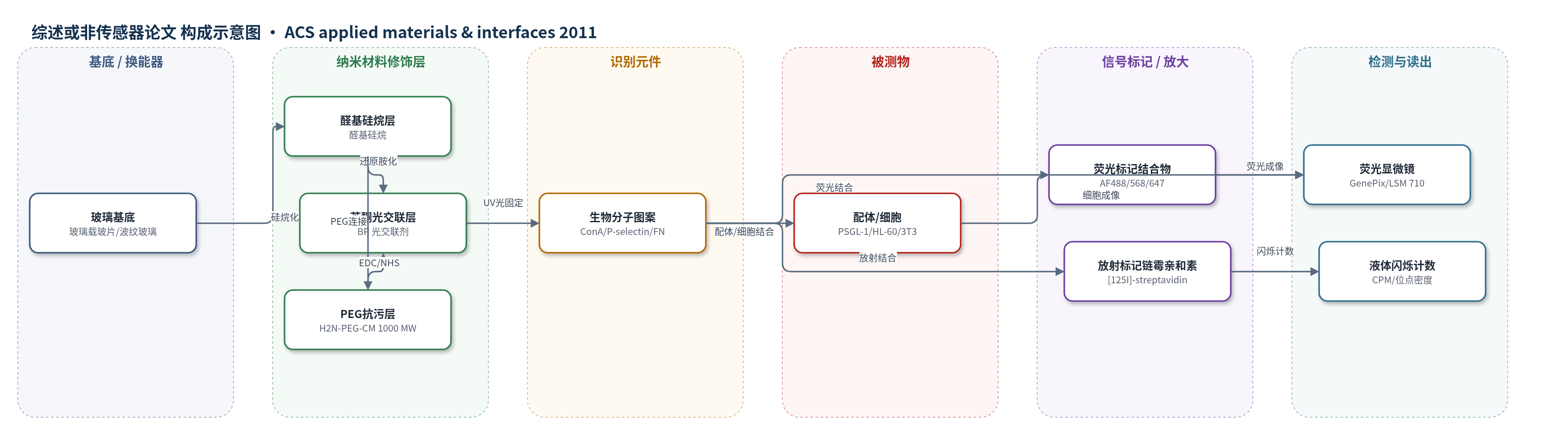
点击图片查看大图 · 依据论文自动绘制
传感器类型
综述或非传感器论文
检测对象
无明确分析物(非传感检测);表面固定/识别对象:ConA-biotin、P-selectin、fibronectin (FN)、mannan;样品基质:PBS、细胞培养液、玻璃基底
检测原理
该方法以苯酮(BP)修饰玻璃为光化学界面。UV(350–365 nm)激发 BP 发生 n→π* 跃迁,生成三重态双自由基,通过质子抽取/自由基重组在表面与溶液生物分子间形成 C–C 键,实现共价固定。曝光时间、光强和生物分子浓度决定固定位点密度,因此可通过改变曝光时间调节表面配体密度。固定后的 ConA-biotin、P-selectin、FN 或 mannan 可被荧光标记结合伙伴或 [125I]-streptavidin 识别;荧光强度或放射性 CPM 与表面固定密度呈正相关,从而将光图案化密度转换为可定量信号。细胞表面受体(如 PSGL-1、整合素)也可识别固定蛋白,实现选择性粘附。
检测灵敏度
原文未报告 LOD、线性范围、灵敏度斜率或相关系数。
效应效果
未报告 RSD、回收率或与 ELISA/HPLC/qPCR 的直接对比。接触角:硅烷化 44.8±1.9°,BP 修饰 53.4±1.1°,蛋白固定后降约 20°;BP-PEG 32.1±4.2°,BP 后 41.8±6.1°。AFM RMS 粗糙度由 352 pm 增至 775 pm,仅亚纳米变化。ConA-biotin 位点密度随 UV 曝光增加,最高约 200 molecules/μm2,荧光强度与 [125I]-streptavidin CPM 可相关转换。P-selectin 特异性结合 PSGL-1 并选择性捕获 HL-60 细胞;FN 图案诱导 3T3 选择性粘附,PEG 背景降低非特异结合。波纹玻璃(100 μm 宽、35 μm 深)上图案强度无明显损失,适用于复杂形貌生物界面。
传感器的构成
- 基底:玻璃载玻片(glass microscope slides),经 Piranha 清洗,提供平面或刻蚀波纹表面
- 硅烷修饰层:3-(triethoxysilyl)butyl aldehyde 气相沉积,形成醛基单分子层
- 光交联剂层:4-benzoyl benzylamine(BP 光交联剂)经 NaCNBH3 还原胺化或 EDC/NHS 偶联固定,提供 UV 光活化 C-H 插入位点
- PEG 抗污层:H2N-PEG-CM(1000 MW)连接醛基,降低非特异蛋白/细胞结合
- 生物分子图案:ConA-biotin、P-selectin、fibronectin(FN)、mannan,经 350–365 nm UV 共价固定
- 封闭/清洗:BSA/PBS、Tween 20、SDS、低 pH Glycine,去除非特异吸附
- 信号标记:Alexa Fluor 488/568/647 标记链霉亲和素、抗 IgG、ConA-AF488、biotinylated anti-FN + AF568 streptavidin,用于荧光识别
- 定量标记:[125I]-streptavidin 放射免疫分析,用于位点密度定量
中文摘要
用于以空间可控方式呈现生物分子的基底生成方法,是生物传感器系统、微阵列技术、基础生物学研究和生物界面科学中的使能工具。本文采用光控制生物分子在苯酮修饰玻璃基底上的直接共价固定,创建生物分子图案。作者生成了按顺序图案化多达三种不同生物分子的基底,并展示了波纹基底上的生物分子光图案化。通过优化底层单分子层化学引入聚乙二醇,使粘附细胞能够粘附到图案化细胞外基质蛋白上。基底经接触角测量、原子力显微镜和免疫荧光显微镜表征。重要的是,采用放射免疫分析定量光图案化基底上固定生物分子的位点密度。通过天然配体识别和细胞粘附实验证明,光图案化蛋白保留功能,表明该方法生成的基底适用于探测特定细胞受体-配体相互作用。这种分子通用的光化学图案化方法,是创建同时具有生物化学和形貌变化基底的使能工具,也是许多天然生物界面的重要特征。
英文摘要
Methods for the generation of substratespresenting biomolecules in a spatially controlled manner are enabling tools for applications in biosensor systems, microarray technologies, fundamental biological studies and biointerface science. We have implemented a method to create biomolecular patterns by using light to control the direct covalent immobilization of biomolecules onto benzophenone-modified glass substrates. We have generated substrates presenting up to three different biomolecules patterned in sequence, and demonstrate biomolecular photopatterning on corrugated substrates. The chemistry of the underlying monolayer was optimized to incorporate poly(ethylene glycol) to enable adhesive cell adhesion onto patterned extracellular matrix proteins. Substrates were characterized with contact angle goniometry, AFM, and immunofluorescence microscopy. Importantly, radioimmunoassays were performed to quantify the site density of immobilized biomolecules on photopatterned substrates. Retained function of photopatterned proteins was demonstrated both by native ligand recognition and cell adhesion to photopatterned substrates, revealing that substrates generated with this method are suitable for probing specific cell receptor-ligand interactions. This molecularly general photochemical patterning method is an enabling tool for the creation of substrates presenting both biochemical and topographical variation, which is an important feature of many native biointerfaces.