组成图示
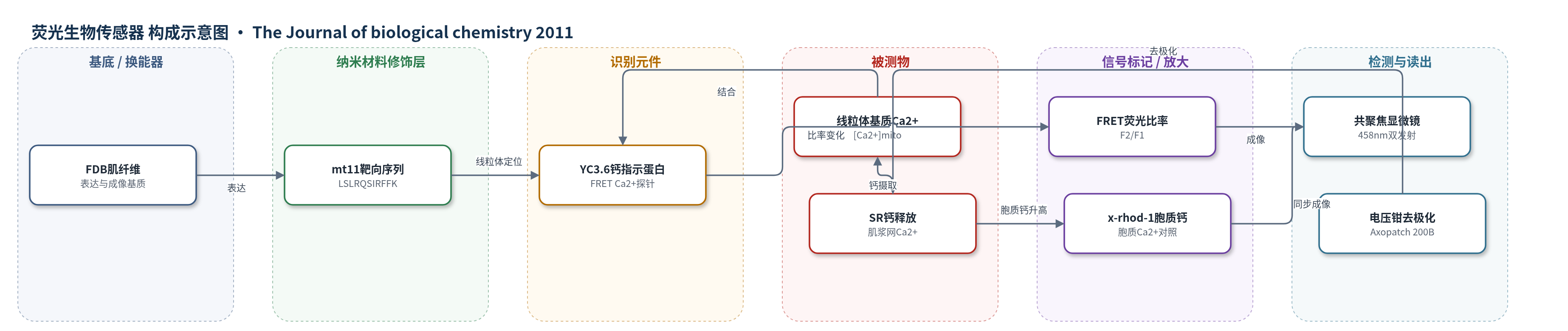
点击图片查看大图 · 依据论文自动绘制
传感器类型
荧光生物传感器
检测对象
线粒体基质钙离子(mitochondrial Ca2+, [Ca2+]mito);同步监测胞质钙离子(cytosolic Ca2+)。样品基质:骨骼肌纤维(FDB,ALS G93A 小鼠)活体细胞内。
检测原理
mt11-YC3.6 由线粒体靶向序列 mt11 与 FRET 钙指示蛋白 YC3.6 融合而成,经电穿孔表达于 FDB 骨骼肌纤维并定位至线粒体。YC3.6 的 Ca2+ 结合引起 458 nm 激发下 F1(470–520 nm)与 F2(520–580 nm)荧光反向变化,计算 F2/F1 比率即可反映线粒体基质 [Ca2+]mito。全细胞电压钳施加去极化脉冲,触发肌浆网(SR)Ca2+ 释放;胞质 Ca2+ 升高后,线粒体通过 Ca2+ 单向转运体(mCU)摄取 Ca2+,使 mt11-YC3.6 比率升高。若用 BAPTA 螯合胞质 Ca2+ 或 Ru360 阻断 mCU,线粒体摄取下降,比率变化减弱,同时胞质 Ca2+ 瞬变升高。因此信号随线粒体钙浓度和摄取能力变化,可定量评估线粒体对 E-C 偶联钙清除的贡献。
检测灵敏度
原文未报告 LOD、线性范围、灵敏度斜率或 R^2。
效应效果
mt11-YC3.6 与 TMRE 共定位,显示良好线粒体靶向性;BAPTA 抑制线粒体 Ca2+ 摄取后信号降低,说明信号来自线粒体。其体内校准动态范围 Rmax/Rmin = 5.7,优于早期 2mtYC2。G93A 肌纤维中,缺陷区胞质 Ca2+ 瞬变升高 10.1 ± 1.7%(n = 8,p < 0.001),线粒体 Ca2+ 摄取降低 14.6 ± 1.9%(n = 4,p = 0.002)。15 mM BAPTA 消除区域差异(1.78 ± 0.39 对 1.79 ± 0.39,p = 0.943)。定量分析显示线粒体峰值清除贡献 18.6 ± 2.4%,峰值流入 4.1 ± 1.0 μM/ms。作者认为该传感器可直接记录骨骼肌线粒体钙动态,为 ALS 机制研究提供工具。
传感器的构成
- 表达基质:FDB 骨骼肌纤维(flexor digitorum brevis),作为 mt11-YC3.6 表达与电压钳成像的活体基质。
- 线粒体靶向层:mt11 序列(LSLRQSIRFFK,源自细胞色素氧化酶亚基 IV),将 YC3.6 定位至线粒体。
- 识别/传感元件:YC3.6 FRET 钙指示蛋白,Ca2+ 结合引起 458 nm 激发下 F1/F2 反向荧光变化。
- 信号标记物:无外源标记,YC3.6 自身 FRET 荧光作为 Ca2+ 信号。
- 辅助识别/对照探针:TMRE(tetramethylrhodamine ethyl ester)标记线粒体膜电位;x-rhod-1 或 fluo-4 监测胞质 Ca2+。
- 读出系统:Leica SP2-AOBS 共聚焦显微镜,458 nm 激发,F1 470–520 nm、F2 520–580 nm,计算 F2/F1 比率。
- 刺激/换能条件:全细胞电压钳(Axopatch 200B)施加去极化脉冲,触发肌浆网(SR)Ca2+ 释放。
中文摘要
肌浆网与线粒体在细胞内 Ca2+ 信号控制中的偶联缺陷被认为参与神经肌肉疾病进展。我们此前发现肌萎缩侧索硬化(ALS)小鼠模型骨骼肌中线粒体功能呈节段性丧失,并伴随肌浆网 Ca2+ 释放活性升高且失控。该局部线粒体缺陷使研究者可在同一肌纤维内比较线粒体正常与缺陷区域,从而评估线粒体在兴奋-收缩(E-C)偶联中清除 Ca2+ 的贡献。本研究发现,膜去极化诱发的 Ca2+ 瞬变在线粒体缺陷区幅度增加约 10%。应用快速 Ca2+ 螯合剂 BAPTA 降低线粒体 Ca2+ 摄取后,上述区域差异消失。利用在 ALS 肌纤维中表达的线粒体靶向 Ca2+ 生物传感器 mt11-YC3.6,我们监测了电压诱导 Ca2+ 释放过程中线粒体 Ca2+ 水平的动态变化,并检测到线粒体缺陷区 Ca2+ 摄取降低,与胞质 Ca2+ 瞬变升高相对应。本研究直接证明线粒体在骨骼肌 E-C 偶联中塑造胞质 Ca2+ 信号的重要性,并确立该机制功能障碍可能参与 ALS 神经肌肉退变。
英文摘要
Defective coupling between sarcoplasmic reticulum and mitochondria during control of intracellular Ca(2+) signaling has been implicated in the progression of neuromuscular diseases. Our previous study showed that skeletal muscles derived from an amyotrophic lateral sclerosis (ALS) mouse model displayed segmental loss of mitochondrial function that was coupled with elevated and uncontrolled sarcoplasmic reticulum Ca(2+) release activity. The localized mitochondrial defect in the ALS muscle allows for examination of the mitochondrial contribution to Ca(2+) removal during excitation-contraction coupling by comparing Ca(2+) transients in regions with normal and defective mitochondria in the same muscle fiber. Here we show that Ca(2+) transients elicited by membrane depolarization in fiber segments with defective mitochondria display an ~10% increased amplitude. These regional differences in Ca(2+) transients were abolished by the application of 1,2-bis(O-aminophenoxy)ethane-N,N,N',N'-tetraacetic acid, a fast Ca(2+) chelator that reduces mitochondrial Ca(2+) uptake. Using a mitochondria-targeted Ca(2+) biosensor (mt11-YC3.6) expressed in ALS muscle fibers, we monitored the dynamic change of mitochondrial Ca(2+) levels during voltage-induced Ca(2+) release and detected a reduced Ca(2+) uptake by mitochondria in the fiber segment with defective mitochondria, which mirrored the elevated Ca(2+) transients in the cytosol. Our study constitutes a direct demonstration of the importance of mitochondria in shaping the cytosolic Ca(2+) signaling in skeletal muscle during excitation-contraction coupling and establishes that malfunction of this mechanism may contribute to neuromuscular degeneration in ALS.