组成图示
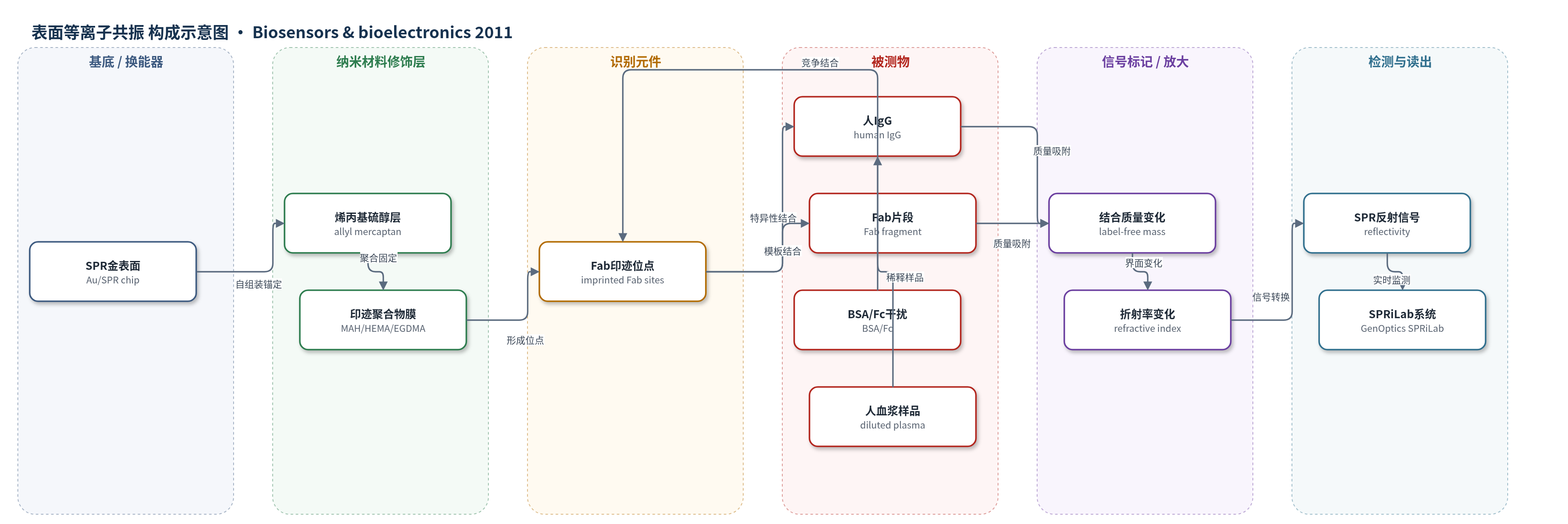
点击图片查看大图 · 依据论文自动绘制
传感器类型
表面等离子共振(SPR)生物传感器
检测对象
人免疫球蛋白G(human IgG)、Fab片段(Fab fragment);样品基质:磷酸缓冲水溶液、稀释人血浆
检测原理
该传感器采用无标记SPR换能机制。SPR芯片金表面经烯丙基硫醇修饰后,以MAH为功能单体、HEMA为基体单体、EGDMA为交联剂,在Fab片段模板存在下光聚合形成分子印迹聚合物纳米膜;洗脱模板后留下与Fab区域互补的印迹识别位点。当人IgG或Fab片段流经芯片时,IgG通过其Fab区与印迹位点特异性结合,结合事件使金表面附近生物质量增加,引起局部折射率变化。SPR仪实时监测反射率(reflectivity)随结合/解离的变化,信号强度随分析物浓度升高而增大。结合过程可用Langmuir单分子层模型描述,1 M NaCl可洗脱再生。该方法无需荧光或酶标记,直接通过质量-折射率传感实现实时定量。
检测灵敏度
LOD: 56 ng/mL;浓度范围: 0.02–0.50 mg/mL(IgG水溶液)、2–15 mg/mL(Fab水溶液);ELISA对比范围: 0.02–1 mg/mL;R^2 = 0.9624(Langmuir拟合);线性回归常数: 99.49%(Fab)、94.1%/96.8%(IgG两段)、99.1%(ELISA对比);斜率: 2.3801(ELISA对比曲线)
效应效果
该SPR芯片对BSA和Fc无特异响应,对Fab和IgG产生特异性响应;在BSA/Fc竞争条件下仍保持对模板Fab和目标IgG的高亲和。选择性系数显示,对Fab相对BSA和Fc分别为2.9306和1.9024,对IgG相对BSA和Fc分别为21.0032和13.6338,表明IgG因分子量更大且含两个Fab区而响应更强。人血浆样品稀释1/800至1/20,000后均可检测,20,000倍稀释(约0.64 μg/mL)仍有响应。与ELISA结果在0.02–1 mg/mL范围内线性一致,曲线为y = 2.3801x + 0.9317,线性回归常数99.1%。作者认为该无标记分子印迹SPR芯片可作为水溶液和复杂血浆样品中IgG实时检测的替代方法。
传感器的构成
- 基底/换能器:SPR芯片金表面(Au/SPR chip),光学换能基底,产生表面等离子共振反射信号
- 锚定修饰层:烯丙基硫醇(allyl mercaptan),在金表面形成可聚合锚定层,用于固定聚合前驱体
- 印迹聚合物纳米膜:N-甲基丙烯酰基-L-组氨酸甲酯(MAH)、2-羟乙基甲基丙烯酸酯(HEMA)和乙二醇二甲基丙烯酸酯(EGDMA)共聚物,形成亲水生物相容识别膜
- 识别元件:Fab片段印迹位点(imprinted Fab sites),去除模板后特异性识别IgG的Fab区域
- 信号标记物:无标记(label-free),结合质量直接改变界面折射率
中文摘要
本研究制备了基于Fab片段印迹的表面等离子共振(SPR)芯片,用于实时检测人免疫球蛋白G(IgG)。首先以烯丙基硫醇修饰SPR芯片金表面,为聚合前驱体提供锚定位点;随后通过木瓜蛋白酶消化IgG并经FPLC/Protein A柱收集Fab片段。将Fab片段与含组氨酸功能单体N-甲基丙烯酰基-L-组氨酸甲酯(MAH)复合,在2-羟乙基甲基丙烯酸酯(HEMA)和乙二醇二甲基丙烯酸酯(EGDMA)存在下于芯片表面光聚合形成分子印迹聚合物纳米膜,并用1 M NaCl磷酸缓冲液洗脱模板。接触角、AFM和FTIR表征表明膜表面亲水且粗糙度增加。实时检测中,芯片对Fab和IgG均呈浓度依赖响应,动力学与等温数据符合Langmuir模型,检出限为56 ng/mL。选择性实验显示芯片对BSA和Fc无特异响应,对IgG/Fab具有较高选择性;人血浆稀释样品检测结果与ELISA结果线性一致,表明该无标记SPR传感器可用于水溶液及复杂生物样品中IgG的实时检测。
英文摘要
F(ab) fragments imprinted surface plasmon resonance (SPR) chip was prepared for the real-time detection of human immunoglobulin G (IgG). In order to attach polymerization precursor on SPR chip, the SPR chip surface was modified with allyl mercaptan. F(ab) fragments of the IgG molecules were prepared by papain digestion procedure and collected by fast protein liquid chromatography (FPLC) system using Hi-Trap_r Protein A FF column. The collected F(ab) fragments were complexed with histidine containing specific monomer, N-methacryloyl-l-histidine methyl ester (MAH). Molecular imprinted polymeric nanofilm was prepared on SPR chip in the presence of ethylene glycol dimethacrylate and 2-hydroxyethylmethacrylate. The template molecules, F(ab) fragments, were removed from the polymeric nanofilm using 1M NaCl solution (pH: 7.4, phosphate buffer system). The molecular imprinted SPR chip was characterized by contact angle, atomic force microscopy and Fourier transform infrared spectroscopy. By the real-time IgG detection studies carried out using aqueous IgG solutions in different concentrations, the kinetics and isotherm parameters of the molecular imprinted SPR chip-IgG system were calculated. To show selectivity and specificity of the molecular imprinted SPR chip, competitive kinetic analyses were performed using bovine serum albumin (BSA), IgG, F(ab) and F(c) fragments in singular and competitive manner. As last step, IgG detection studies from human plasma were performed and the measured IgG concentrations were well matched with the results determined by enzyme-linked immunosorbent assay (ELISA). The results obtained with the molecular imprinted SPR chip were well fitted to Langmuir isotherm and the detection limit was found as 56 ng/mL. In the light of the results, we can conclude that the proposed molecular imprinted SPR chip can detect IgG molecules from both aqueous solutions and complex natural samples.