组成图示
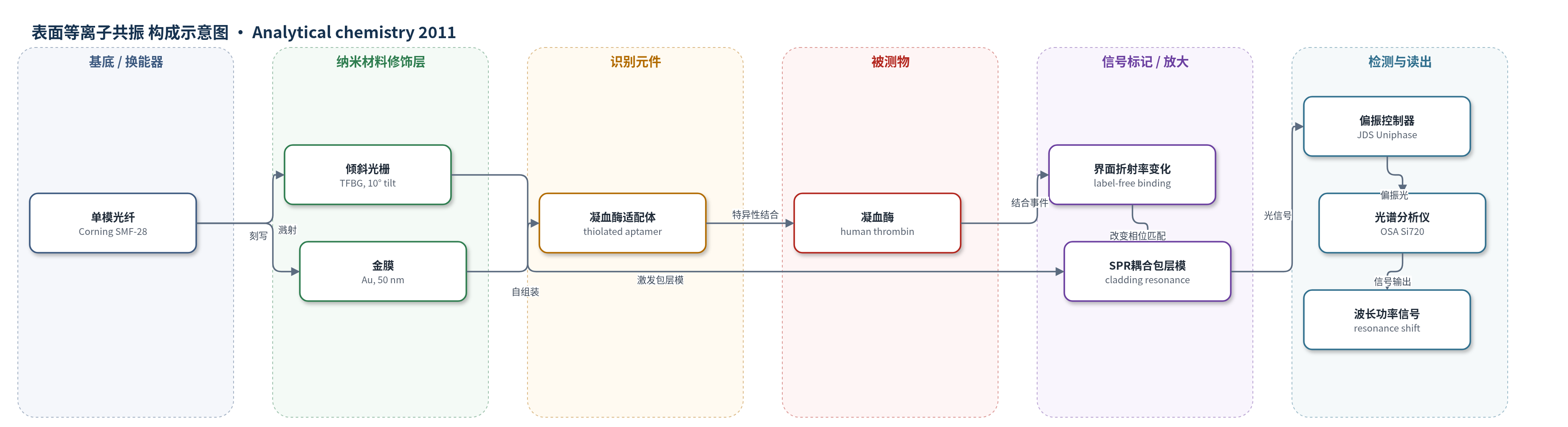
点击图片查看大图 · 依据论文自动绘制
传感器类型
表面等离子共振(SPR)生物传感器
检测对象
凝血酶(thrombin,human thrombin);样品基质:蛋白缓冲液、50%牛血清/Tris缓冲液。
检测原理
传感器以标准单模光纤为基底,纤芯内倾斜光纤布拉格光栅(TFBG)将导波耦合到多个包层模;光纤包层外溅射50 nm金膜,当包层模满足相位匹配条件时激发表面等离子共振(SPR)。5'-巯基凝血酶适配体通过硫醇自组装固定于金表面,作为识别元件。凝血酶与适配体特异性结合后,金膜附近纳米尺度内的局部折射率增加,改变 SPR 相位匹配条件,使最敏感的 SPR 耦合包层共振峰发生波长移动或功率变化。光学传感分析仪追踪该共振峰,信号随凝血酶吸附量增加而增强。该方案为无标记检测,不依赖酶或荧光放大;TFBG 的芯模共振还可用于消除温度交叉灵敏度。
检测灵敏度
LOD: 22.6 nM
效应效果
该传感器可实时监测适配体自组装,并在10 min内区分0.1 μM凝血酶与蛋白缓冲液;100 min后特异性与非特异性信号差异最大。对照实验显示5 μM BSA和5 μM胃蛋白酶仅产生非特异性响应。在含30 mg/mL非特异性蛋白的50%牛血清中,5 μM(0.18 mg/mL)凝血酶仍可被检测,血清与血清加凝血酶的SPR差约0.07 dB,与缓冲液中5 μM凝血酶差0.074 dB相近。凝血酶实验信噪比2.11–194.51,适配体实验8.58–36.97;LOD为22.6 nM,Kd约40±1 nM,与文献20–200 nM一致。共聚焦和AFM证实适配体及蛋白结合。
传感器的构成
- 基底/换能器:标准单模光纤 Corning SMF-28,纤芯内刻写倾斜光纤布拉格光栅 TFBG(倾角10°),用于激发包层模并耦合 SPR。
- 金属修饰层:金(Au)薄膜,真空溅射沉积,厚度50 nm,提供 SPR 激发界面。
- 识别元件:5'-巯基己基修饰凝血酶适配体(thrombin aptamer,HS-(CH2)6-5'-GGT TGG TGT GGT TGG-3'),通过硫醇自组装固定于金表面,特异性识别凝血酶。
- 信号标记物:无标记(label-free),不添加荧光、酶或纳米标记物,结合事件直接改变界面折射率。
- 读出系统:偏振控制器与光学传感分析仪(OSA,Micron Optics Si720),在1520–1570 nm追踪最敏感 SPR 耦合包层共振峰。
中文摘要
基于光纤的表面等离子共振(SPR)生物传感器可作为监测生物分子相互作用的低成本、易实现平台,用于原位甚至可能体内监测。本研究采用在纤芯中刻写倾斜光纤布拉格光栅(TFBG)的标准单模光纤,在宽外部折射率范围内激发光纤表面 SPR,且对温度交叉灵敏度低,不破坏光纤结构。该无标记生物识别方案通过金涂层光纤表面固定适配体实现,适配体为可与目标物高特异性结合的合成 DNA 序列。研究实时监测了适配体在光纤表面的功能化过程,并检测了缓冲液和血清中不同浓度凝血酶的存在。结果还表明,该 SPR 生物传感器可用于评估解离常数(Kd),所得结合常数与文献报道值一致。
英文摘要
Surface plasmon resonance (SPR) biosensors prepared using optical fibers can be used as a cost-effective and relatively simple-to-implement alternative to well established biosensor platforms for monitoring biomolecular interactions in situ or possibly in vivo. The fiber biosensor presented in this study utilizes an in-fiber tilted Bragg grating to excite the SPR on the surface of the sensor over a large range of external medium refractive indices, with minimal cross-sensitivity to temperature and without compromising the structural integrity of the fiber. The label-free biorecognition scheme used demonstrates that the sensor relies on the functionalization of the gold-coated fiber with aptamers, synthetic DNA sequences that bind with high specificity to a given target. In addition to monitoring the functionalization of the fiber by the aptamers in real-time, the results also show how the fiber biosensor can detect the presence of the aptamer's target, in various concentrations of thrombin in buffer and serum solutions. The findings also show how the SPR biosensor can be used to evaluate the dissociation constant (K(d)), as the binding constant agrees with values already reported in the literature.