组成图示
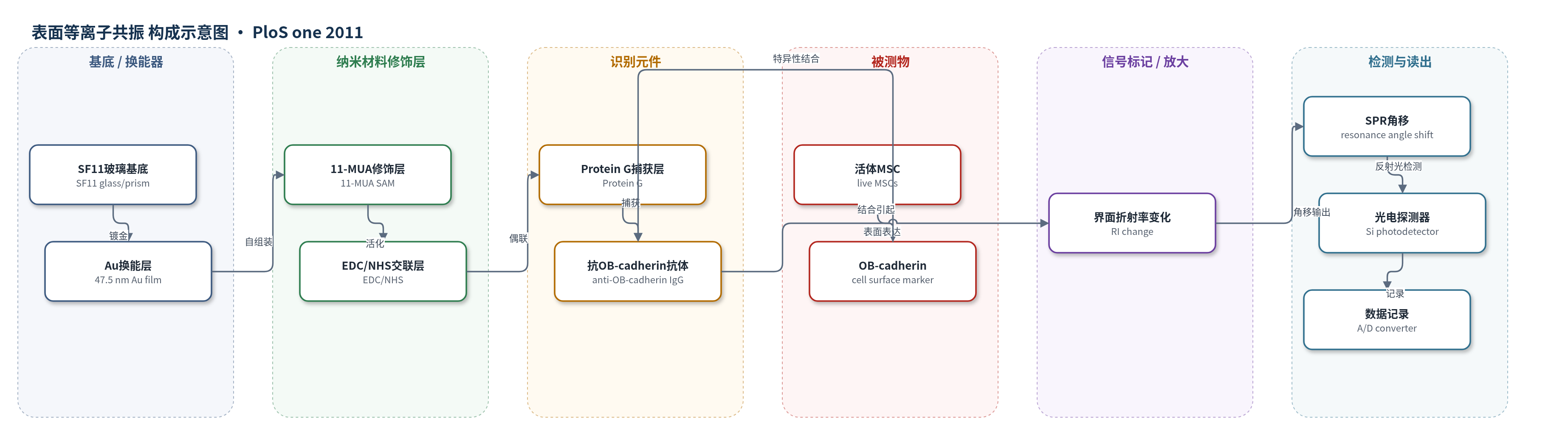
点击图片查看大图 · 依据论文自动绘制
传感器类型
表面等离子共振(SPR)生物传感器
检测对象
活体间充质干细胞(live mesenchymal stem cells, MSCs)的成骨分化程度;样品基质:PBS活细胞悬液
检测原理
该SPR生物传感器以Au薄膜为换能界面,经11-MUA自组装、EDC/NHS活化、Protein G固定和抗OB-cadherin抗体偶联实现功能化,并用BSA封闭。活体MSC在PBS中流过细胞室,细胞表面OB-cadherin与固定抗体发生特异性抗原-抗体结合。成骨诱导时间越长,MSC表面OB-cadherin表达越高,被捕获的细胞越多,Au界面附近质量与折射率增加,导致SPR共振角移增大。系统以无抗体芯片、mouse IgG芯片、SaOS2阳性细胞和Hep3B阴性细胞扣除非特异结合,并将MSC角移与SaOS2角移归一化,从而定量反映OB-cadherin表达水平。该过程无标记、无酶放大,信号随成骨分化程度增加而线性增强。
检测灵敏度
原文未报告具体LOD、线性范围、灵敏度斜率或相关系数数值;仅报告成骨诱导时间与折射角移差呈线性关系且相关系数很高。
效应效果
系统以无抗体Au芯片和mouse IgG芯片作为非特异结合对照,SaOS2为OB-cadherin阳性对照、Hep3B为阴性对照,可区分阳性细胞与未诱导MSC。SPR在成骨诱导第0–6天即可定量区分OB-cadherin表达差异,而Western blot在第6天前不可测、第12天前信号较弱,仅趋势一致。细胞室温度稳定在37°C,波动小于0.1°C;成骨诱导15天后MSC与SaOS2的OB-cadherin表达接近。作者认为该SPR方法可活细胞、无标记、实时、定量评估成骨成熟,优于传统Western blot,并可用于肿瘤侵袭性预测和其他干细胞分化监测。
传感器的构成
- 基底/换能器:SF11玻璃片与47.5 nm Au薄膜,作为SPR换能基底,激发表面等离子极化激元并反射光信号
- 自组装修饰层:1 mM 11-MUA(11-巯基十一酸)在75%乙醇中修饰Au表面,提供羧基用于后续共价偶联
- 交联活化层:2 mM:5 mM EDC/NHS活化11-MUA羧基,实现Protein G与Au表面共价连接
- 捕获层:0.005 μg/mL Protein G固定于Au表面,用于捕获抗体
- 识别元件:0.05 μg/mL mouse IgG anti-human OB-cadherin(抗人OB-cadherin小鼠IgG)特异性结合细胞表面OB-cadherin
- 封闭剂:1 mM BSA封闭非特异性结合位点
- 流体/温控模块:带流体通道的细胞室与TE cooler,维持37°C并灌注PBS/DI水,使活细胞流过芯片并洗涤未结合细胞
- 光学读出模块:He-Ne激光(632.8 nm)、偏振器、分束器、硅光电探测器、电机旋转台与A/D转换器,测量反射光强随入射角变化并输出共振角移
中文摘要
表面等离子共振(SPR)生物传感器因对测试对象折射率变化高度敏感,已成为实时动态分析分子结合亲和力的有用工具。传统分子生物学方法评估细胞分化通常需要细胞裂解或固定,难以在活细胞中进行,且需要一定数量细胞以获得足够蛋白或信使RNA用于检测。为克服该局限,作者开发了一种基于SPR的生物传感装置,利用特定抗原-抗体结合引起的细胞表面光学性质差异,实时检测活细胞中的细胞分化。本研究将该SPR系统用于评估间充质干细胞(MSCs)的成骨分化。成骨分化过程中上调表达的OB-cadherin作为靶标,通过在传感芯片表面偶联抗OB-cadherin抗体实现识别。结果显示,成骨诱导时间与折射角移差之间呈线性关系,且相关系数很高。总之,该SPR生物传感器可在活细胞、无标记、无需破细胞条件下快速准确判定MSC成骨成熟,有望促进生物医学研究和医学诊断。
英文摘要
Surface plasmon resonance (SPR) biosensors have been recognized as a useful tool and widely used for real-time dynamic analysis of molecular binding affinity because of its high sensitivity to the change of the refractive index of tested objects. The conventional methods in molecular biology to evaluate cell differentiation require cell lysis or fixation, which make investigation in live cells difficult. In addition, a certain amount of cells are needed in order to obtain adequate protein or messenger ribonucleic acid for various assays. To overcome this limitation, we developed a unique SPR-based biosensing apparatus for real-time detection of cell differentiation in live cells according to the differences of optical properties of the cell surface caused by specific antigen-antibody binding. In this study, we reported the application of this SPR-based system to evaluate the osteogenic differentiation of mesenchymal stem cells (MSCs). OB-cadherin expression, which is up-regulated during osteogenic differentiation, was targeted under our SPR system by conjugating antibodies against OB-cadherin on the surface of the object. A linear relationship between the duration of osteogenic induction and the difference in refractive angle shift with very high correlation coefficient was observed. To sum up, the SPR system and the protocol reported in this study can rapidly and accurately define osteogenic maturation of MSCs in a live cell and label-free manner with no need of cell breakage. This SPR biosensor will facilitate future advances in a vast array of fields in biomedical research and medical diagnosis.