组成图示
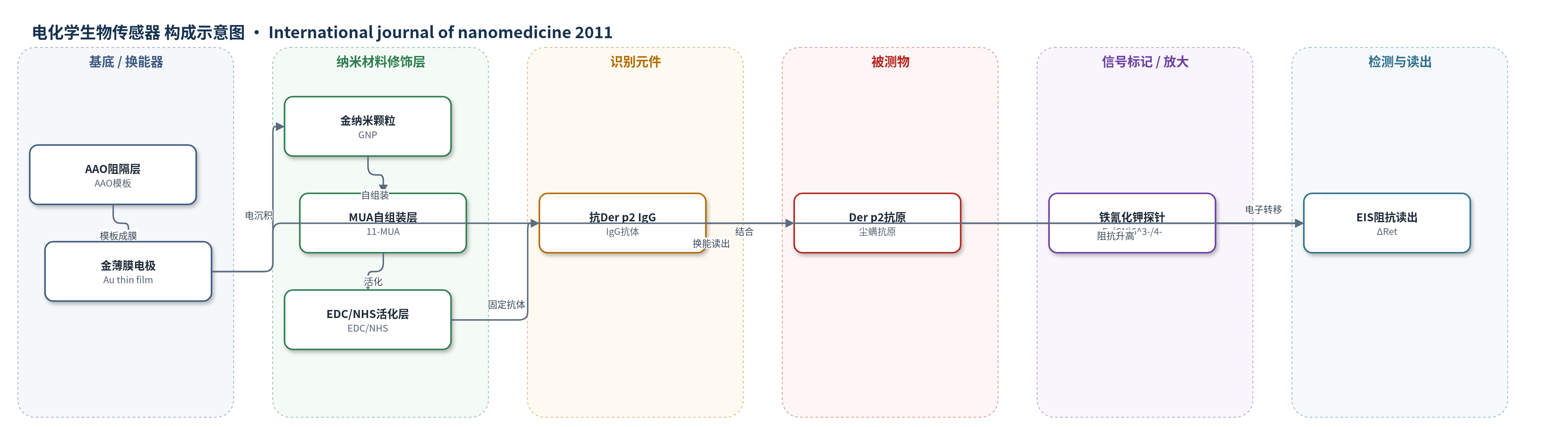
点击图片查看大图 · 依据论文自动绘制
传感器类型
电化学生物传感器
检测对象
尘螨抗原 Der p2(Dermatophagoides pteronyssinus group 2 allergen);样品基质:Der p2标准溶液/缓冲液(EIS使用100 mM MOPS,pH 6.0)
检测原理
该传感器以AAO阻隔层为模板,在其上溅射30 nm金薄膜并退火,再于-0.8 V电沉积约10 nm金纳米颗粒,形成三维纳米结构工作电极。11-MUA在金表面自组装形成羧基锚定层,经EDC/NHS活化后与尘螨单克隆IgG的氨基共价结合,1% BSA封闭非特异位点。检测时,Der p2抗原与固定IgG特异性结合,使电极界面疏水性和空间位阻增加,阻碍Fe(CN)6^3-/4-氧化还原探针在电极表面的电子转移。EIS在0 V、5 mV、0.1–100 kHz下测量,用Randles等效电路拟合,电荷转移电阻Ret随Der p2浓度升高而增大;以ΔRet对Der p2对数浓度作图呈线性,从而实现无标记定量检测。
检测灵敏度
LOD: 1 pg/mL;动态范围: 5 µg/mL;R^2 = 0.99
效应效果
该传感器在EIS检测中表现出良好线性,R^2=0.99,检出限1 pg/mL,动态范围达5 µg/mL;各浓度重复3次,标准偏差均小于5%,重现性较好。与平面金薄膜相比,AAO/Au/GNP电极有效面积增大4.18倍,Der p2-EGFP共聚焦荧光分析显示其荧光面积百分比由5.12%提高到22.76%,荧光强度由13,409提高到59,670,粘附量约提高4.45倍,说明三维金纳米结构显著增加抗体结合位点并增强信号。论文未报告选择性、抗干扰、稳定性或实际样品回收率,也未与ELISA等方法直接对比,但作者认为该传感器可用于溶液中稀有分子的快速检测。
传感器的构成
- AAO模板层:阳极氧化铝(AAO)阻隔层,纳米孔约60 nm、厚度约50 µm,提供三维模板与支撑
- 金薄膜电极:30 nm Au薄膜,射频磁控溅射沉积并120°C退火,作为工作电极/换能器
- 金纳米颗粒修饰层:约10 nm Au纳米颗粒(GNP),由HAuCl4在-0.8 V电化学沉积,增大有效面积与结合位点
- MUA自组装层:5 mM 11-巯基十一烷酸(11-MUA)自组装单分子层,提供羧基锚定
- EDC/NHS活化层:EDC/NHS混合液活化羧基,用于抗体共价固定
- 识别元件:尘螨单克隆抗体(IgG,10 µg/mL),特异性识别Der p2
- 封闭层:1% BSA溶液,封闭非特异性结合位点
- 信号介质:5 mM Fe(CN)6^4-/Fe(CN)6^3-(100 mM MOPS,pH 6.0),用于EIS电子转移阻抗测量
中文摘要
第2组尘螨过敏原Der p2可激活呼吸道上皮细胞上的天然Toll样受体(TLRs),从而加重呼吸道疾病。本研究提出一种基于三维传感元件且均匀沉积金纳米颗粒的高灵敏纳米结构生物传感器,用于检测尘螨抗原Der p2。该传感器的阻隔层由阳极氧化铝(AAO)膜构成,并作为模板。无需还原剂和稳定剂的简单电化学沉积即可在阻隔层表面均匀合成金纳米颗粒;纳米颗粒的尺寸和分布密度可通过电化学沉积过程中的施加电位良好控制。随后,通过11-巯基十一烷酸(11-MUA)和EDC/NHS自组装单分子层方法固定尘螨单克隆抗体(IgG),用于Der p2检测。采用电化学阻抗谱(EIS)分析考察该三维金纳米颗粒纳米结构生物传感器的检测限,结果为1 pg/mL,动态范围为5 µg/mL。该传感器有望用于溶液中稀有分子的快速检测。
英文摘要
The group 2 allergen, Der p2, has been reported to activate innate toll-like receptors (TLRs) on respiratory epithelial cells and thus aggravate respiratory diseases. In this study, a highly sensitive nanostructured biosensor based on a 3D sensing element with uniformly deposited gold nanoparticles is proposed for the detection of the dust mite antigen Der p2. The barrier layer comprises an anodic aluminum oxide (AAO) film which is used as the template in this highly sensitive nanostructured biosensor. Simple electrochemical deposition without reducing agent and stabilizer was enough to uniformly synthesize gold nanoparticles on the surface of the barrier layer. The size and the distribution density of the nanoparticles can be well controlled by the applied potential during electrochemical deposition. Following this procedure, the dust mite monoclonal antibodies (IgG) were then immobilized through the 11-MUA (11-mercaptoundecanoic acid), (1-ethyl-3-(3-dimethyl-aminopropyl)-carbodiimide)/(N-hydroxysuccinimide) self-assembled monolayer approach for the dust mite antigen Der p2 detection. The detection limit of the proposed 3D gold nanoparticle-based nanostructured biosensor was examined using electrochemical impedance spectroscopy analysis and found to be 1 pg/mL. The dynamic range was found to be 5 μg/mL. The proposed nanostructured biosensor would be useful for fast detection of rare molecules in a solution.