组成图示
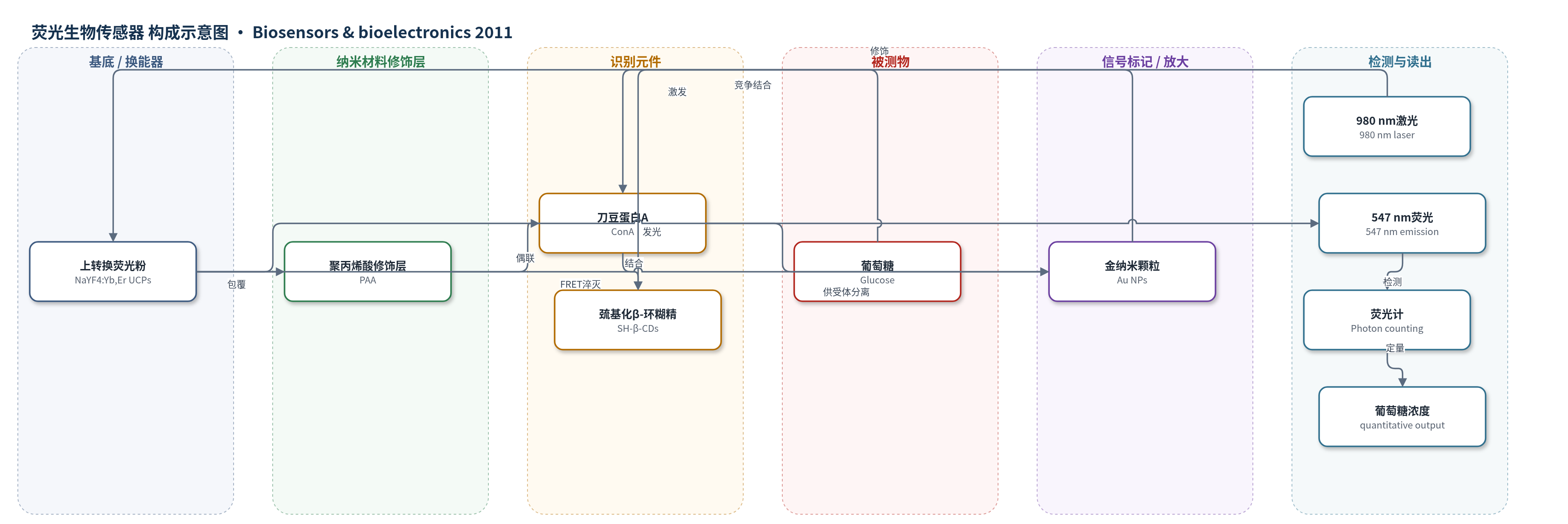
点击图片查看大图 · 依据论文自动绘制
传感器类型
荧光生物传感器
检测对象
葡萄糖(glucose);样品基质:人血清(human serum,含经葡萄糖氧化酶预处理去除内源葡萄糖的血清及真实人血清)
检测原理
该传感器利用上转换荧光共振能量转移(UC-FRET)实现葡萄糖检测。NaYF4:Yb,Er上转换荧光粉(UCPs)经PAA修饰后偶联刀豆蛋白A(ConA),作为能量供体;金纳米颗粒(Au NPs)表面修饰巯基化β-环糊精(SH-β-CDs),作为能量受体。ConA与SH-β-CDs特异性结合,使UCPs与Au NPs距离进入FRET有效范围,UCPs的547 nm上转换荧光被Au NPs非辐射淬灭。加入葡萄糖后,葡萄糖与SH-β-CDs竞争ConA结合位点,UCPs–ConA–SH-β-CDs-Au复合体解离,供受体距离增大,FRET效率下降,UCPs荧光恢复。980 nm激光激发下,547 nm荧光强度随葡萄糖浓度升高而增强,从而实现定量检测。
检测灵敏度
LOD: 0.043 μM(水缓冲液);线性范围: 0.4 μM–10 μM(水缓冲液);LOD: 0.065 μM(人血清);线性范围: 0.4 μM–10 μM(人血清)
效应效果
该传感器对葡萄糖具有良好选择性:在10倍浓度干扰糖及血清正常水平金属离子、氨基酸、蛋白存在下,荧光相对偏差均在±6%以内。预处理人血清中线性范围为0.4–10 μM,检出限0.065 μM,与缓冲液体系相近,说明UCPs近红外激发可有效抵抗血清背景自荧光和散射干扰。真实人血清样品检测中,UC-FRET法结果与己糖激酶法一致,相对标准偏差为2.7%–6.2%,加标回收率为96.5%–105%。作者认为该方法无需光学标记酶反应,可直接用于复杂生物基质中葡萄糖检测,具有临床应用潜力。
传感器的构成
- 荧光供体/信号标记物:NaYF4:Yb,Er上转换荧光粉(UCPs),980 nm激发下547 nm发光,作为UC-FRET能量供体
- 表面修饰层:聚丙烯酸(PAA)包覆UCPs,提供羧基、水溶性和偶联位点
- 偶联试剂:EDC·HCl与Sulfo-NHS活化PAA-UCPs羧基,实现ConA共价偶联
- 识别元件:刀豆蛋白A(ConA)偶联到PAA-UCPs,特异性结合葡萄糖/β-环糊精
- 能量受体纳米颗粒:金纳米颗粒(Au NPs),吸收UCPs发射并淬灭其荧光,作为FRET受体
- 受体识别配体:巯基化β-环糊精(SH-β-CDs)通过Au–S键修饰Au NPs,并与ConA结合拉近供受体距离
中文摘要
本文报道了一种基于上转换荧光共振能量转移(UC-FRET)的新型葡萄糖传感器。将共价标记刀豆蛋白A(ConA)的上转换荧光粉(UCPs,NaYF4:Yb,Er)作为能量供体,将巯基化β-环糊精(SH-β-CDs)功能化的金纳米颗粒作为能量受体。由于ConA与SH-β-CDs特异性结合,供体与受体被拉近,金纳米颗粒淬灭UCPs荧光。当葡萄糖存在时,葡萄糖与SH-β-CDs竞争ConA结合位点,使UCPs–ConA–SH-β-CDs-Au复合体解离,供体与受体分离,UCPs荧光随葡萄糖浓度恢复。在水缓冲液中,UCPs荧光强度在0.4–10 μM范围内与葡萄糖浓度成正比,检出限为0.043 μM;在预处理人血清基质中获得相同线性范围,检出限略高为0.065 μM。该传感器用于真实人血清样品检测,结果与经典己糖激酶法一致,表明UC-FRET生物传感器可借助近红外激发直接检测血清中葡萄糖,避免光学干扰,有望用于复杂生物样品诊断。
英文摘要
In this work, a new glucose sensor based on up-converting fluorescence resonance energy transfer (UC-FRET) was developed. Up-converting phosphors (UCPs, NaYF(4): Yb, Er), which were covalently labeled with Concanavalin A (ConA), were used as the energy donor with thiolated β-cyclodextrins (SH-β-CDs) functionalized gold nanoparticles as the energy acceptor. Due to the combination between ConA and SH-β-CDs, the energy donor and the acceptor were brought to close proximity, resulting in the quenching of the fluorescence of UCPs by gold nanoparticles. In the presence of glucose which competed with SH-β-CDs towards the binding sites of ConA, the biosensor (UCPs-ConA-SH-β-CDs-Au) was decomposed and the energy donor was separated from the acceptor. Therefore, the fluorescence of UCPs was restored dependent on the concentration of glucose. The increase of UCPs fluorescence intensity was proportional to glucose concentration within the range from 0.4 μM to 10μM in aqueous buffer, with a limit of detection (LOD) of 0.043 μM. A same linear range of glucose concentration was obtained in a human serum matrix (which was pretreated and thus contained no glucose) with a slightly higher LOD (0.065 μM). The glucose sensor was applied to real human serum samples with the results consistent with that of a classic hexokinase (HK) method, indicating that the UC-FRET biosensor was competent for directly sensing glucose in serum samples without optical interference, which benefited from the near infrared (NIR) excitation nature of UCPs. The results of this work suggested that the UC-FRET technique could be a promising alternative for detecting biomolecules in complex biological sample matrixes for diagnostic purposes.