组成图示
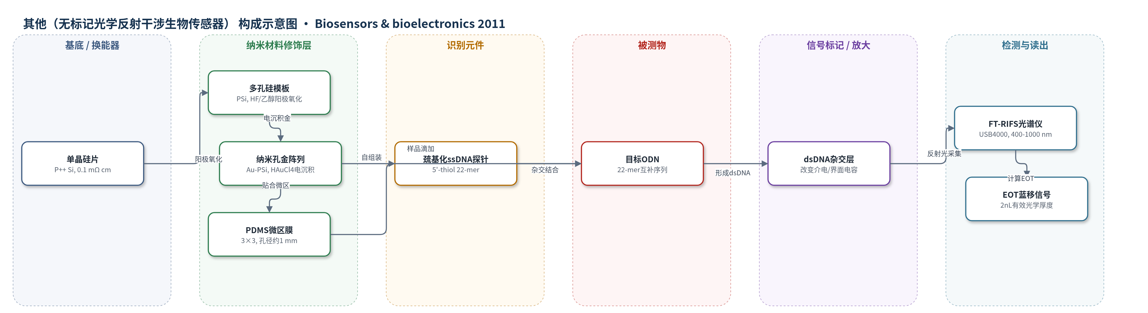
点击图片查看大图 · 依据论文自动绘制
传感器类型
其他(无标记光学反射干涉生物传感器)
检测对象
目标寡脱氧核苷酸(target oligodeoxynucleotides, ODNs,22-mer DNA);样品基质:Tris-HCl-EDTA缓冲液(pH 7.4)
检测原理
该传感器为无标记光学检测。5'-巯基化ssDNA探针通过Au–S键自组装于Au–PSi纳米孔内壁,目标ODN与探针互补杂交形成dsDNA。杂交后双链结构更刚性并沿孔壁伸展,使DNA层厚度增加;按串联电容模型,界面电容C1下降,纳米孔层有效介电常数和有效折射率随之改变。由于Au折射率低于Si和空气,Au–PSi本身已使EOT蓝移;DNA固定与杂交进一步降低有效折射率,导致EOT(2nL)持续蓝移。FT-RIFS通过反射干涉峰位置变化读出EOT位移,其大小随ODN浓度增加而增大。纳米孔结构还可尺寸过滤大分子DNA,提高选择性。
检测灵敏度
LOD: 约10−14 mol L−1;基线噪声: EOT变化约5 nm
效应效果
该传感器选择性与稳定性良好。非互补ssDNA(10−5 mol L−1)处理后EOT变化与空白基本一致(n=5,P=0.95),非特异性吸附可忽略。纳米孔可阻止含互补片段的大DNA分子进入传感层,降低大分子干扰;含巯基小分子可能因与金表面高亲和而干扰。Au–PSi芯片在PBS中保存3个月后,对同一ODN浓度信号无明显变化,表明金层保护PSi并提高化学稳定性。高比表面使高浓度ODN未饱和,动态范围增大。作者认为其可用于ODN药物发现及ODN与目标DNA结合检测,并有望作为多孔电极用于电化学生物传感。
传感器的构成
- 基底/换能器:单晶硅片(P++,0.1 mΩ cm)经HF/乙醇电化学阳极氧化形成多孔硅(PSi)模板,提供纳米孔阵列与光学干涉层。
- 纳米材料修饰层:在PSi孔道内壁电沉积金纳米层(HAuCl4,5 mA,10 min),形成PSi模板纳米孔金阵列(Au-PSi),保留多孔结构并提高化学稳定性。
- 识别元件:5'-巯基化22-mer ssDNA探针(5'-thiol-CTA CAG GTG AAG GTG GAA TGG T)经Au-S键自组装于Au-PSi孔壁,捕获互补ODN。
- 样品结合层:目标ODN(22-mer互补序列 GAT GTC CAC TTC CAC CTT ACC A)与探针杂交形成dsDNA,改变孔壁介电常数与界面电容。
- 传感区定义层:图案化聚二甲基硅氧烷(PDMS)膜(3×3,孔径约1.0 mm)贴合芯片,定义传感区并支持重复检测。
- 信号读出层:傅里叶变换反射干涉光谱(FT-RIFS)/USB 4000光纤光谱仪,监测400–1000 nm反射干涉并计算有效光学厚度(EOT)蓝移。
中文摘要
本研究报道了一种基于多孔硅(PSi)模板制备有序纳米孔金阵列的方法,用于无标记光学检测寡脱氧核苷酸(ODN)。在低电解电流和低浓度氯金酸(HAuCl4)溶液中,可在PSi表面电沉积金纳米层,所得PSi模板纳米孔金(Au–PSi)阵列复制了PSi的纳米孔结构并保留其光学性质。傅里叶变换反射干涉光谱显示,由于金膜折射率较低,Au–PSi的特征有效光学厚度(EOT)发生蓝移。随后,通过特定序列单链DNA(ssDNA)在Au–PSi表面的自组装构建光学DNA生物传感器。ssDNA的固定及其与目标ODN的杂交均持续引起EOT蓝移,从而建立了EOT位移与ODN浓度之间的关系。作者用电磁理论和电化学阻抗谱(EIS)对DNA杂交引起的光学响应机制进行了定性解释。在基线噪声对应EOT变化约5 nm时,目标ODN最低检测限约为10−14 mol L−1。该Au–PSi光学生物传感器有望用于发现新的ODN药物,因为它能够检测ODN与目标DNA之间的结合事件。
英文摘要
Interest in using nanoporous materials for sensing applications has increased. The present study reports a method of preparing well-ordered nanoporous gold arrays using a porous silicon (PSi) template. Gold nanolayer could be electrodeposited on the surface of the PSi template at low electrolysis currents in low concentration of chloroauric acid (HAuCl(4)) solution. Surface morphology characterizations and optical measurements revealed that a PSi-templated nanoporous gold (Au-PSi) array well replicated the nanoporous structure and retained the optical properties of PSi. Fourier transform reflectometric interference spectra showed that a characteristic blue-shifted effective optical thickness (EOT) was observed due to the low refractive index of the gold film. An optical DNA biosensor was then fabricated via the self-assembly of single-stranded DNA (ssDNA) with a specific sequence on the surface of Au-PSi. The attachment of ssDNA and its hybridization with target oligonucleotides (ODNs) persistently caused the blue shift of the EOT. Consequently, a relationship between the EOT shift and the ODN concentration was established. The mechanism of the optical response caused by DNA hybridization on the Au-PSi surface was qualitatively explained by the electromagnetic theory and electrochemical impedance spectroscopy (EIS). The lowest detection limit for target ODNs was estimated at around 10(-14) mol L(-1), when the baseline noise, a variation in the value of EOT is around 5 nm. The fabricated Au-PSi based optical biosensor has potential use in the discovery of new ODN drugs because it will be able to detect the binding event between ODNs and the target DNA.