组成图示
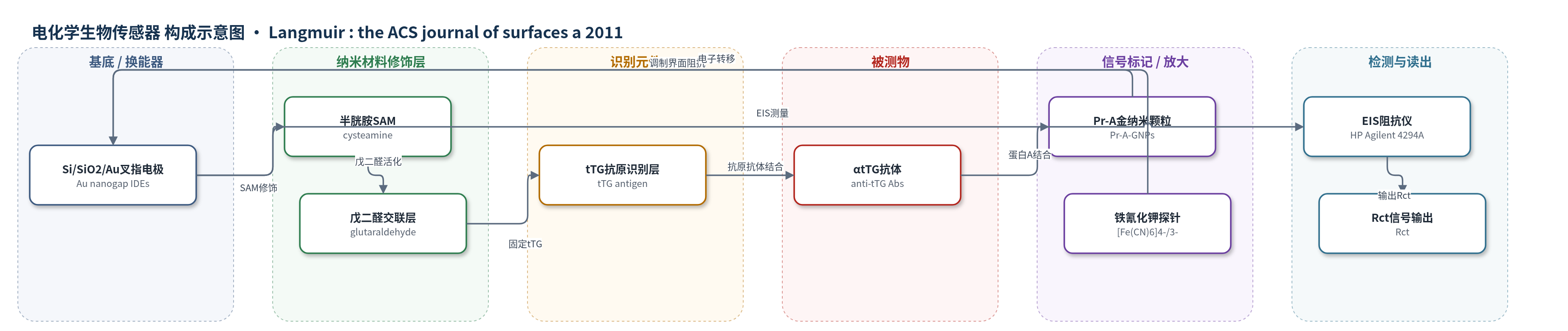
点击图片查看大图 · 依据论文自动绘制
传感器类型
电化学生物传感器
检测对象
抗组织转谷氨酰胺酶抗体(anti-tissue transglutaminase antibodies, αtTG-Abs);样品基质:PBST稀释的山羊抗tTG血清/血清
检测原理
传感器以Au纳米间隙叉指电极为换能器,表面经半胱胺SAM、戊二醛固定tTG抗原。血清中αtTG-Abs与tTG特异性结合,形成界面生物层;随后Pr-A-GNPs通过蛋白A结合IgG Fc区,将金纳米颗粒引入电极表面。检测采用法拉第EIS,以K4[Fe(CN)6]/K3[Fe(CN)6]为氧化还原探针。抗体结合改变电极/溶液界面的电荷转移过程:无标记时生物层阻碍电子转移,Rct随抗体量增加而增大;优化稀释的Pr-A-GNP进一步放大界面阻抗变化,使Rct变化幅度与捕获抗体量成正比,从而实现灵敏度增强。
检测灵敏度
动态检测范围(估算): 30 pM–30 nM;IC50(血清稀释度): ELISA 0.15×10^-4,无标记 0.98×10^-4,GNP标记 2.7×10^-4;灵敏度斜率(归一化信号/稀释度): 无标记 2.63,GNP标记 9.15,ELISA 9.49;Rct变化: 无标记 490–1850 Ω(3.75倍),GNP标记 550–5110 Ω(9.3倍);ELISA OD450: 0.307–2.7(8.8倍)
效应效果
传感器通过非特异抗原OVA对照验证了检测特异性。重现性方面,芯片ELISA标准差为平均值的3–5%,阻抗生物传感器为12–18%,作者指出阻抗测量易受温度波动影响,实验在72°F室温控制下进行。与芯片ELISA相比,无标记检测信号仅变化3.75倍,灵敏度低于ELISA;Pr-A-GNP标记后信号变化达9.3倍,归一化灵敏度斜率由2.63提高到9.15,接近ELISA的9.49,且最低检测限与ELISA相当。作者认为该平台有望用于即时电化学免疫检测,减少ELISA所需集中式精密仪器。
传感器的构成
- 基底/换能器电极:650 μm Si片、1 μm SiO2层及金叉指电极(Au nanogap IDEs),提供纳米间隙电场与法拉第EIS换能
- 自组装单分子层:半胱胺(cysteamine)SAM,在Au表面形成氨基功能化层
- 交联活化层:戊二醛(glutaraldehyde,2.5% PBS),活化SAM氨基并共价固定抗原
- 识别元件:组织转谷氨酰胺酶(tTG,50 μg/mL)抗原,捕获血清中αtTG-Abs
- 封闭层:乙醇胺(ethanolamine,1%)中和未反应基团,聚乙烯吡咯烷酮(PVP,1%)封闭非特异结合
- 信号标记物:蛋白A偶联胶体金纳米颗粒(Pr-A-GNPs,15–20 nm),结合IgG Fc区并增强阻抗信号
- 氧化还原探针:K4[Fe(CN)6]/K3[Fe(CN)6](各2.5 mM,10 mM PBS),提供法拉第电子转移并反映界面阻抗变化
中文摘要
本文报道了一种基于纳米间隙叉指电极阵列(IDEA)的阻抗免疫生物传感器。首先通过电场与电流密度有限元模拟优化纳米间隙叉指电极几何参数,结果表明电极间隙(G)对电场分布和电流密度的影响大于宽度(W)和高度(H)。随后制备多种器件,并采用法拉第电化学阻抗谱(EIS)评价其对电化学环境的灵敏度。优化后的纳米间隙IDE被用于无标记亲和检测抗组织转谷氨酰胺酶抗体(αtTG-Abs),该抗体是乳糜泻的血清生物标志物。结果显示,无标记检测灵敏度低于芯片ELISA。进一步引入蛋白A偶联金纳米颗粒(Pr-A-GNPs)作为信号标记,使换能器灵敏度较无标记检测提高350%。研究证实纳米间隙IDE适用于EIS无标记和GNP标记免疫检测,GNP标记后免疫生物传感器检测灵敏度与ELISA相当。
英文摘要
Interdigitated electrode (IDE) arrays with nanometer-scale gaps have been utilized to enhance the sensitivity of affinity-based detection. The geometry of nanogap IDEs was first optimized on the basis of simulations of the electric field and current density. It was determined that the gap (G) between the electrodes was the most important geometric parameter in determining the distribution and strength of the electric field and the current density compared to the width (W) and height (H) of the IDEs. Several devices were materialized and analyzed for their sensitivity to the electrochemical environment using faradic electrochemical impedance spectroscopy (EIS) as the detection technique. Nanogap optimized IDEs were then employed as biosensors for the label-free, affinity-based detection of antitissue transglutaminase antibodies (αtTG-Abs), a biomarker for the detection of autoimmune disorder celiac sprue, triggered by ingesting gluten. The label-free biosensor assay was found to be less sensitive compared to on-chip ELISA. Gold nanoparticles (GNPs) were then employed to improve the sensitivity of the nanogap IDE-based biosensor. With GNPs, the transducer sensitivity increased by 350% over that of label-free detection. The suitability of nanogap IDEs as biosensor transducers for EIS in label-free and GNP-labeled formats was established. The immunobiosensor assay detection sensitivity with the GNPs was found comparable to ELISA.