组成图示
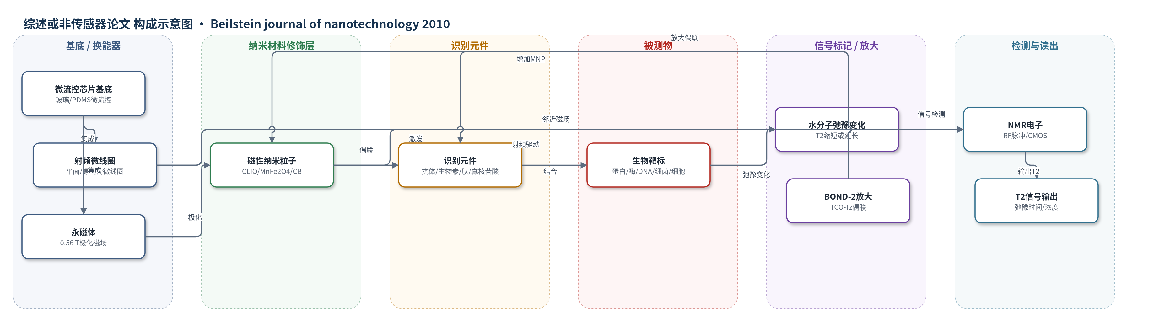
点击图片查看大图 · 依据论文自动绘制
传感器类型
综述或非传感器论文
检测对象
DNA/mRNA、蛋白生物标志物(GFP、CA-125、VEGF、α-fetoprotein、β-HCG、telomerase)、小分子/药物(folate、glucose、D-phenylalanine)、酶活性(caspase-3、trypsin、renin、MMP-2、MPO)、细菌(S. aureus、BCG/MTB)、病毒(HSV-1、Adenovirus-5)、肿瘤细胞(BT474、小鼠异种移植FNA);样品基质:缓冲液、血液、痰液、细胞悬液、细针穿刺液。
检测原理
DMR以靶向磁性纳米粒子(MNP)作为邻近传感器。MNP在外部磁场中产生局部磁场,增加水分子周围磁场不均匀性;水质子扩散经过MNP表面时,自旋相干进动被扰动,纵向T1和横向T2弛豫时间缩短,通常以T2作为读出。对小分子靶标,采用磁弛豫切换(MRSw):正向切换中靶标作为交联剂使MNP聚集成簇,有效横截面积增大,T2缩短;反向切换中酶切或竞争结合使预形成簇解聚,T2延长。对细菌或细胞等大靶标,靶向MNP结合表面标志物后洗涤去除未结合MNP,1/T2与结合MNP数量成正比。BOND-2利用TCO修饰抗体与Tz修饰MNP的快速无催化Diels–Alder环加成,提高MNP结合价态并放大信号;微线圈与CPMG序列测量T2变化。
检测灵敏度
LOD: GFP低达低飞摩尔范围;S. aureus为每微升样品数个菌落形成单位(CFUs);BCG用CLIO约100 CFUs、用高弛豫率cannonballs约6 CFUs;内置过滤后约1 CFU;痰液样品1 ml中至少20 CFUs;细胞检测约2个细胞(1 µL样品体积);线性范围: S. aureus动态范围超过三个数量级;avidin四个数量级动态范围
效应效果
综述报道的DMR系统具有良好选择性:GFP检测中BSA对照不引起T2变化,caspase-3检测中加入特异性抑制剂可阻止簇解聚。MnFe2O4的r2达420 s−1·mM−1[metal],比CLIO(约50 s−1·mM−1[Fe])高8倍以上;cannonball饱和磁化强度为139 emu·g−1[Fe]。与结核传统培养及抗酸杆菌涂片相比,DMR可在30分钟内从1 ml痰液中检出约20 CFUs。细胞检测与流式细胞术和Western blot相关良好,所需细胞更少,时间少于15分钟;多标志物分析可提高恶性诊断准确性。微型化μNMR支持微升级样品并行检测,适合即时检测。
传感器的构成
- 换能器基底:玻璃基底或聚合物微流控芯片,承载微线圈与样品通道
- 射频微线圈:平面微线圈阵列或螺线管微线圈(solenoidal microcoil),用于射频激发与NMR信号接收
- 磁场源:小型永磁体(portable permanent magnet,0.56 T),提供极化磁场
- 微流控网络:PDMS/聚合物微流控通道,用于样品进样、混合、分配及膜过滤清洗
- 磁性纳米粒子:CLIO(Fe3O4/γ-Fe2O3核、葡聚糖涂层、伯胺功能化)、MnFe2O4(DMSA涂层)或Fe@铁氧体cannonball,作为邻近传感器与信号标记
- 识别元件:抗体(anti-GFP、anti-Her2、anti-EGFR、anti-EpCAM、anti-BCG)、生物素/亲和素、肽探针(DEVD/GDEVDG)、寡核苷酸探针或万古霉素,赋予分子特异性
- 信号放大偶联:BOND-2中TCO修饰抗体与Tz修饰MNP经Diels–Alder环加成偶联,提高MNP结合价态
- NMR电子读出:分立射频芯片或CMOS IC,生成RF脉冲序列(CPMG)并放大处理NMR信号
中文摘要
快速、准确地测量生物样品中的蛋白生物标志物、病原体和细胞,可为早期疾病诊断、治疗监测和个体化医疗提供重要信息。由于生物样品本身磁化率极低,利用磁性纳米粒子(MNPs)进行生物传感不仅能提高灵敏度,还可减少样品前处理。本综述聚焦于基于磁共振效应的体外生物分子与细胞检测,即诊断磁共振(DMR)。DMR 将靶向 MNPs 作为邻近传感器,通过改变其周围水分子的自旋-自旋弛豫时间(T2)产生信号。近年来,随着更优 MNPs、微型化 NMR 检测器和新型偶联方法的发展,DMR 对 DNA/mRNA、蛋白、小分子/药物、细菌和肿瘤细胞等靶标的检测限显著改善,并可在微升级样品中实现并行、高灵敏测量,有望成为便携式、低成本、高效的生物医学检测平台。
英文摘要
Rapid and accurate measurements of protein biomarkers, pathogens and cells in biological samples could provide useful information for early disease diagnosis, treatment monitoring, and design of personalized medicine. In general, biological samples have only negligible magnetic susceptibility. Thus, using magnetic nanoparticles for biosensing not only enhances sensitivity but also effectively reduces sample preparation needs. This review focuses on the use of magnetic nanoparticles for in vitro detection of biomolecules and cells based on magnetic resonance effects. This detection platform, termed diagnostic magnetic resonance (DMR), exploits magnetic nanoparticles as proximity sensors, which modulate the spin-spin relaxation time of water molecules surrounding molecularly-targeted nanoparticles. By developing more effective magnetic nanoparticle biosensors, DMR detection limits for various target moieties have been considerably improved over the last few years. Already, a library of magnetic nanoparticles has been developed, in which a wide range of targets, including DNA/mRNA, proteins, small molecules/drugs, bacteria, and tumor cells, have been quantified. More recently, the capabilities of DMR technology have been further advanced with new developments such as miniaturized nuclear magnetic resonance detectors, better magnetic nanoparticles and novel conjugational methods. These developments have enabled parallel and sensitive measurements to be made from small volume samples. Thus, the DMR technology is a highly attractive platform for portable, low-cost, and efficient biomolecular detection within a biomedical setting.