组成图示
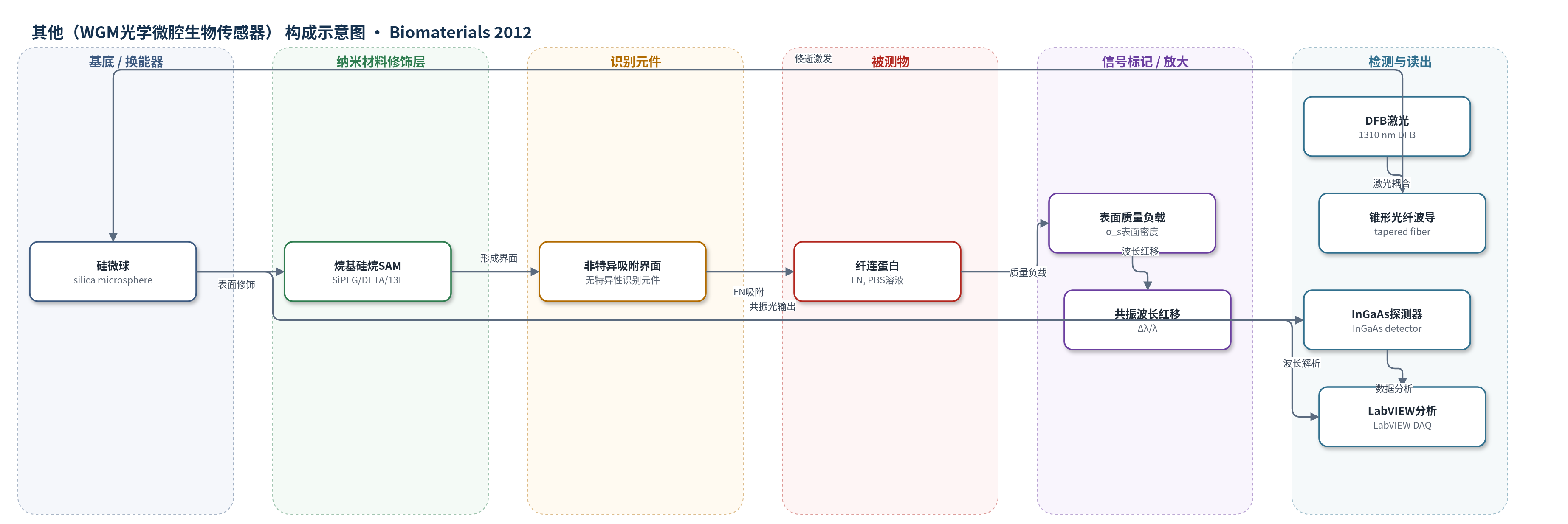
点击图片查看大图 · 依据论文自动绘制
传感器类型
其他(WGM光学微腔生物传感器)
检测对象
纤连蛋白(fibronectin, FN);样品基质:50 mM PBS(pH 7.4)牛血浆纤连蛋白溶液,以及用于细胞培养的硅烷修饰盖玻片/微球表面
检测原理
该传感器基于回音壁模式(WGM)光学微腔。近红外DFB激光经锥形光纤波导倏逝耦合进入二氧化硅微球,形成高Q共振。当PBS中的纤连蛋白(FN)非特异性吸附到SiPEG、DETA或13F烷基硅烷SAM表面时,微球表面质量/折射率负载增加,等效半径和边界条件改变,导致共振波长发生红移。依据一阶微扰理论,Δλ/λ与吸附表面密度σ_s近似成正比,因此可通过连续监测共振波长变化定量吸附动力学。微流控流动池维持层流,CFD模拟用于校正近表面浓度和传质限制;方法为无标记检测,不依赖酶、荧光或电化学放大,灵敏度主要来自高Q WGM共振和微球表面高比表面积。
检测灵敏度
LOD: ~1 pg/mm2
效应效果
该WGM系统灵敏度约1 pg/mm2,作者称比SPR高约10倍,可在10、1、0.5和0.25 mg/mL FN浓度下无标记定量吸附。10 mg/mL时饱和表面密度为13F 200 ng/cm2、DETA 190 ng/cm2、SiPEG 25 ng/cm2;1 mg/mL时为13F约135 ng/cm2、DETA约137 ng/cm2、SiPEG约14 ng/cm2,低浓度下与烷硫醇SAM文献差异显著。细胞实验显示DETA表面FN生物活性最高:海马活细胞212±102 cells/mm2,肌管35±13;13F表面活细胞少且无肌管,SiPEG表面几乎无细胞存活。作者认为该装置可研究硅烷表面蛋白积累与细胞响应,适用于生物材料评价。
传感器的构成
- 基底/换能器:二氧化硅微球(silica microsphere,半径125–175 μm),作为WGM光学微腔,表面吸附改变有效半径/折射环境并引起共振波长红移
- 表面修饰层:烷基硅烷自组装单层(alkylsilane SAMs,SiPEG、DETA、13F),提供亲水/带电/疏水/全氟等不同表面化学,用于FN非特异吸附
- 识别/捕获层:无特异性生物识别元件,仅依靠FN与SiPEG/DETA/13F SAM之间的非特异性吸附形成表面负载
- 信号标记物:无外源标记物,采用无标记(label-free)检测,FN本身作为质量/折射率负载产生共振波长偏移
- 流体耦合层:锥形光纤波导(tapered optical fiber waveguide,SMF-28e+)与微球倏逝耦合;聚碳酸酯流动池(polycarbonate flow cell)和PDMS垫片维持层流
- 信号读出:DFB激光器(1310 nm)、InGaAs光电探测器、LabVIEW数据采集与分析系统,监测共振波长并计算表面浓度
中文摘要
本研究构建了一种回音壁模式(WGM)生物传感器,用于在其它技术难以达到的低溶液浓度下测量蛋白质在烷基硅烷自组装单层(SAM)上的吸附。传感器以功能化烷基硅烷SAM的二氧化硅微球作为WGM谐振器,并集成于层流微流控流动池中,通过监测微球共振波长变化实现高灵敏度、无标记检测。实验发现,纤连蛋白(FN)可在具有生物学意义的表面密度下吸附,但其吸附动力学和浓度依赖饱和值与以往基于烷硫醇SAM的报道存在显著差异。作者将随机顺序吸附模型和吸附后转变模型拟合实验数据,以解释观察到的动力学。随后在修饰表面上培养胚胎海马神经元和骨骼肌成肌细胞,并用活/死染色评估FN表面的细胞培养适用性,发现不同SAM引起明显不同的细胞响应。WGM生物传感器的高灵敏度、结构简单以及可定量任意稀蛋白吸附的能力,使其成为研究表面蛋白积累及其对细胞功能影响的重要工具,对体内和体外生物材料应用具有重要意义。
英文摘要
A Whispering Gallery Mode (WGM) biosensor was constructed to measure the adsorption of protein onto alkysilane self-assembled monolayers (SAMs) at solution concentrations unattainable with other techniques. The high sensitivity was provided by a WGM resonance excited in a silica microsphere that was functionalized with alkylsilane SAMs and integrated in a microfluidic flow cell under laminar flow conditions. It was found that FN adsorbed at biologically relevant surface densities, however, the adsorption kinetics and concentration dependent saturation values varied significantly from work published utilizing alkanethiol SAMs. Mathematical models were applied to the experimental results to interpret the observed kinetics of FN adsorption. Embryonic hippocampal neurons and skeletal myoblasts were cultured on the modified surfaces, a live--dead assay was used to determine the viability of the FN surfaces for cell culture, and major differences were noted in the biological response to the different SAMs. The high sensitivity and simplicity of the WGM biosensor, combined with its ability to quantify the adsorption of any dilute protein in a label-free assay, establishes the importance of this technology for the study of surface accretion and its effect on cellular function, which can affect biomaterials for both in vivo and in vitro applications.