组成图示
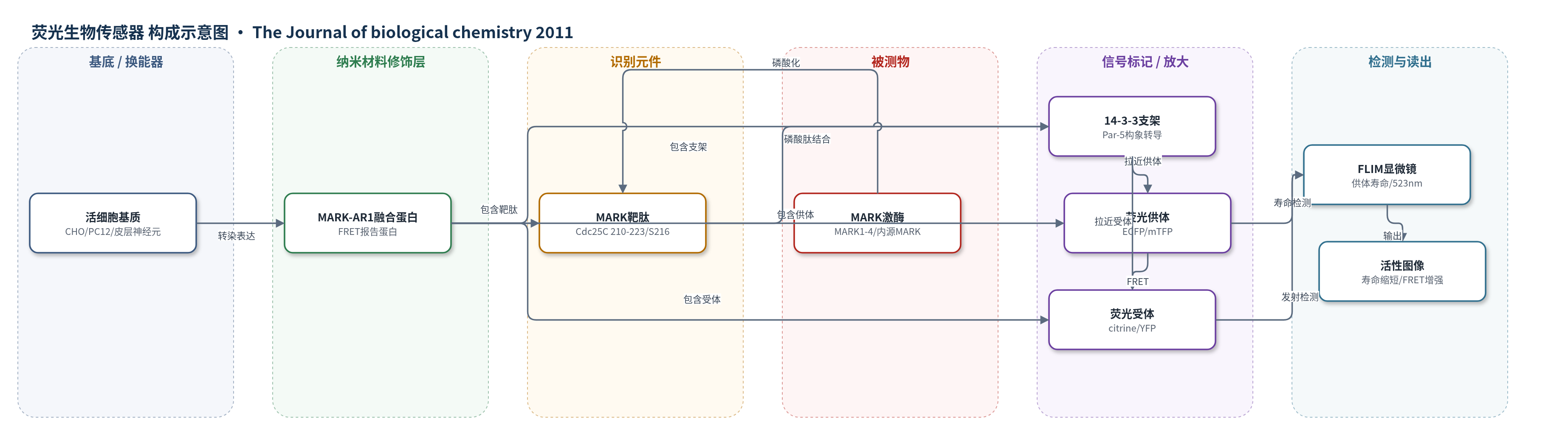
点击图片查看大图 · 依据论文自动绘制
传感器类型
荧光生物传感器
检测对象
MARK/Par-1 激酶活性(MARK/Par-1 kinase activity);样品基质:CHO 细胞、PC12 细胞、原代皮层神经元(活细胞)及体外重组激酶反应体系
检测原理
MARK-AR1 将 Cdc25C 210–223 靶肽(含 Ser-216)连接在 14-3-3 支架蛋白与 ECFP/mTFP、citrine/YFP 之间。内源或外源 MARK 磷酸化靶肽后,磷酸肽进入 14-3-3 结合口袋,使 14-3-3 构象闭合,供体与受体距离缩短,FRET 增强。此时供体荧光寿命缩短,受体 523 nm 发射增强;FLIM 通过记录供体寿命变化实现定量读出。MARK 活性越高,寿命越短、FRET 越强;加入特异性抑制剂后磷酸化受抑,寿命恢复,从而反映激酶活性变化。
检测灵敏度
原文未报告 LOD、线性范围、灵敏度斜率或 R^2。
效应效果
传感器可区分 MARK 与多数 Tau 激酶:除 SAD-B 外,PKA、CaMKII、MAPKAPK2、Chk1/2 不能有效诱导 FRET;26203 和 39621 对 MARK 选择性较高。CHO 中供体寿命由 2.42 ns 降至 2.18 ns;神经元生长锥活性区寿命 1.897±0.007 ns,非磷酸化突变体约 2.2 ns。39621 处理后寿命升至 2.166±0.013 ns,轴突生长速度由 32.553±2.515 μm/h 降至 5.773±1.195 μm/h,可实时监测内源 MARK 活性。
传感器的构成
- 细胞基质:CHO、PC12 或原代皮层神经元,提供内源 MARK 活性与活细胞检测环境
- 荧光供体:ECFP 或 mTFP(teal fluorescent protein),位于融合蛋白 N 端,作为 FRET 供体
- 识别元件:Cdc25C 210–223 肽段(含 Ser-216),作为 MARK 特异性磷酸化靶序列
- 构象转导元件:14-3-3(Par-5)支架蛋白,结合磷酸化靶肽后改变构象并拉近供受体
- 荧光受体:citrine(YFP 稳定变体),位于融合蛋白 C 端,作为 FRET 受体并在 523 nm 发射
- 表达系统:Lipofectamine 2000 转染,将 MARK-AR1 融合蛋白导入活细胞
- 换能读出:FLIM 共聚焦显微镜(Olympus FluoView FV1000、PicoQuant TCSPC),通过供体荧光寿命变化报告 MARK 活性
中文摘要
微管亲和力调节激酶(MARK)/Par-1 家族蛋白激酶在细胞极性建立、细胞周期调控和胞内信号转导中发挥重要作用,其功能紊乱与癌症及脑疾病(如无脑回畸形和阿尔茨海默病)相关。为阐明 MARK 家族激酶的生物学作用,作者筛选特异性抑制剂并开发 MARK 活性生物传感器。对约 18000 个化合物的 ChemBioNet 库进行筛选,获得若干低微摩尔范围具有抑制活性的化合物,它们可在培养细胞和原代神经元中抑制 MARK 活性,判断依据为 MARK 依赖性微管相关蛋白磷酸化及其对微管完整性的影响。其中四个化合物共享 9-oxo-9H-acridin-10-yl 结构,可作为优化抑制效率的先导结构。为检验这些抑制剂,作者构建了细胞内 MARK 活性生物传感器:将 MARK 靶序列连接至 14-3-3 支架蛋白,并与增强青色/蓝绿和黄色荧光蛋白组成 FRET 供受体对。将蓝绿/黄色荧光蛋白传感器转染神经元后,通过荧光寿命成像发现 MARK 在分化神经元的轴突和生长锥中尤为活跃。这些抑制剂可能通过干扰阿尔茨海默病中异常的 Tau 磷酸化而具有治疗潜力。
英文摘要
Protein kinases of the microtubule affinity regulating kinase (MARK)/Par-1 family play important roles in the establishment of cellular polarity, cell cycle control, and intracellular signal transduction. Disturbance of their function is linked to cancer and brain diseases, e.g. lissencephaly and Alzheimer disease. To understand the biological role of MARK family kinases, we searched for specific inhibitors and a biosensor for MARK activity. A screen of the ChemBioNet library containing ~18,000 substances yielded several compounds with inhibitory activity in the low micromolar range and capable of inhibiting MARK activity in cultured cells and primary neurons, as judged by MARK-dependent phosphorylation of microtubule-associated proteins and its consequences for microtubule integrity. Four of the compounds share a 9-oxo-9H-acridin-10-yl structure as a basis that will serve as a lead for optimization of inhibition efficiency. To test these inhibitors, we developed a cellular biosensor for MARK activity based on a MARK target sequence attached to the 14-3-3 scaffold protein and linked to enhanced cyan or teal and yellow fluorescent protein as FRET donor and acceptor pairs. Transfection of the teal/yellow fluorescent protein sensor into neurons and imaging by fluorescence lifetime imaging revealed that MARK was particularly active in the axons and growth cones of differentiating neurons.