组成图示
点击图片查看大图 · 依据论文自动绘制
传感器类型
表面等离子共振(SPR)生物传感器
检测对象
肿瘤细胞系(A549、U-2 OS、Saos-2、CHO-K1、HT29、LS174T、HeLa、T84、MCF12A、MCF-7、LCC1、LCC2),样品基质为细胞培养液中的贴壁细胞
检测原理
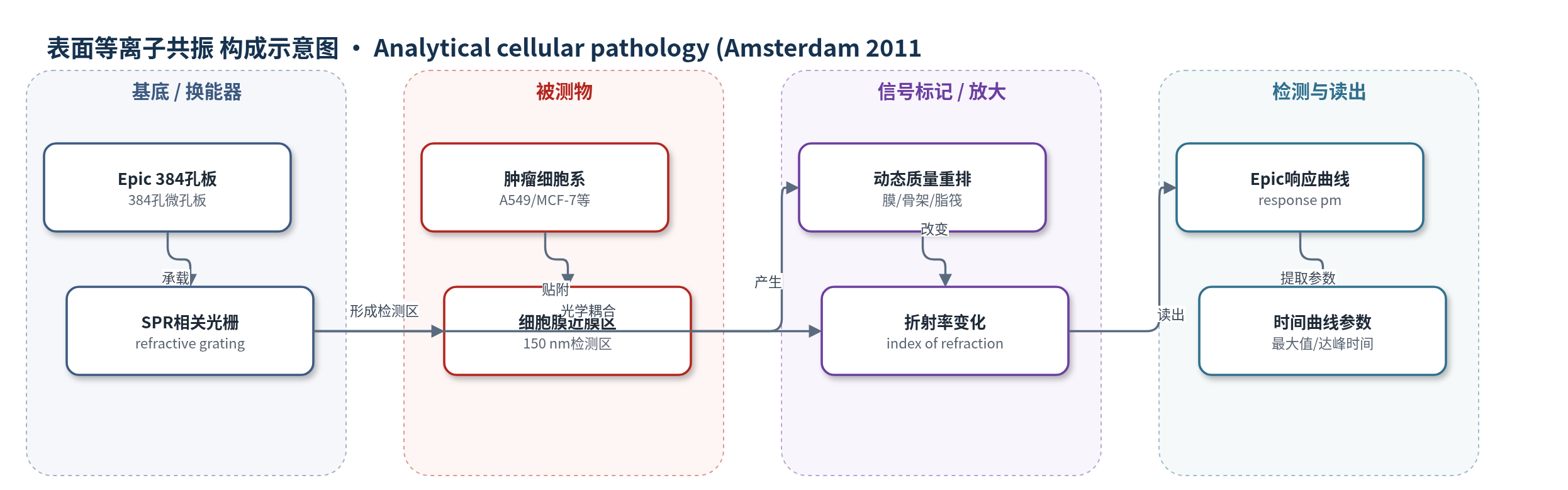
肿瘤细胞贴附于Epic微孔板底部的折射光栅表面,宽带光源激发光栅,通过表面等离子共振相关现象在细胞膜内及近膜约150 nm检测区形成敏感场。细胞膜受体激活、二聚化、内化,细胞骨架重排,脂筏运动,细胞黏附、形状改变以及内吞/胞吐等活动引起局部大分子质量重排,改变该区域折射率。折射率变化被光栅换能器转换为响应值(pm),系统每45 s采集一次,每点为5次扫描平均。不同细胞系因膜组成、骨架动力学和迁移相关活动不同,产生不同时间曲线;无外源标记或酶放大,信号为全细胞整合性生物物理响应。
检测灵敏度
未报告
效应效果
重复孔数据高度可重复,同一细胞系各孔在任意时间点的响应值接近且曲线形状一致;未报告RSD、选择性、抗干扰或回收率。达峰时间从30分钟到3.5小时不等,最大值从最低的LS174T到最高的Saos-2差异明显。接种后开始采集的时间(30分钟至12小时)会影响曲线形状和最大值,但各细胞系保持自身特征;12小时内不存在使所有细胞系产生相同谱的起始点。多数肿瘤类型中,异种移植转移能力更强的细胞最大值较低,但乳腺癌细胞相反:MCF-12A最高,MCF-7低于转移性LCC1/LCC2。达峰时间与转移行为未见相关。作者认为该无标记生物物理谱可辅助研究肿瘤侵袭转移,并需与阻抗等多方法互补。
传感器的构成
- 基底/换能器:384孔Epic微孔板,底部为薄折射光栅(refractive waveguide grating),经宽带光源照明,SPR相关现象形成约150 nm检测区
- 识别/传感对象:肿瘤细胞系(A549、U-2 OS、Saos-2、CHO-K1、HT29、LS174T、HeLa、T84、MCF12A、MCF-7、LCC1、LCC2),贴附于孔底,作为无标记传感对象
- 样品基质:细胞培养液(F-12K、McCoy's 5A、DMEM、IMEM等,含10% FBS及相应补充物),维持细胞活性
- 信号标记物:无外源信号分子(signal-free/label-free),细胞膜及近膜区动态质量重排本身产生折射率变化
- 读出层:Epic光学生物传感器系统,实时采集响应值(pm),每45 s一个数据点,每点为5次扫描平均
中文摘要
尽管肿瘤细胞遗传谱差异显著,但癌细胞共享促进侵袭和转移的表型特征,包括膜相关改变。本研究使用Corning Epic光学生物传感器实时监测源自结肠、骨、宫颈、肺和乳腺癌的肿瘤细胞系中细胞膜内及近膜区的动态质量重排。该方法无需外源信号分子,属于无标记检测。各细胞系在时间过程中呈现独特谱:均先升至最大值,但达峰速率、最大值及峰值后下降速率不同。信号最大值与细胞在异种移植实验中的转移行为存在相关;多数细胞类型中,小鼠体内转移能力更强的细胞时间曲线最大值较低,但乳腺癌细胞呈相反趋势。这些独特谱及至少一个谱特征与转移行为的相关性,表明生物物理肿瘤细胞谱在癌症生物学研究中具有潜在应用价值。
英文摘要
Despite significant differences in genetic profiles, cancer cells share common phenotypic properties, including membrane-associated changes that facilitate invasion and metastasis. The Corning Epic optical biosensor was used to monitor dynamic mass rearrangements within and proximal to the cell membrane in tumor cell lines derived from cancers of the colon, bone, cervix, lung and breast. Data was collected in real time and required no exogenously added signaling moiety (signal-free technology). Cell lines displayed unique profiles over the time-courses: the time-courses all displayed initial signal increases to maximal values, but the rate of increase to those maxima and the value of those maxima were distinct for each cell line. The rate of decline following the maxima also differed among cell lines. There were correlations between the signal maxima and the observed metastatic behavior of the cells in xenograft experiments; for most cell types the cells that were more highly metastatic in mice had lower time-course maxima values, however the reverse was seen in breast cancer cells. The unique profiles of these cell lines and the correlation of at least one profile characteristic with metastatic behavior demonstrate the potential utility of biophysical tumor cell profiling in the study of cancer biology.